Diagnosing Stroke

Finally! A randomized clinical trial evaluating treatments for cerebral venous sinus thrombosis (CVST)

When the topic of stroke is being discussed, for the most part it is the concept of “blockages” developing within in the arteries of the brain – that is, the blood vessels that are carrying oxygen-rich blood *to* the brain to nurture its cells. These blockages can occur from the formation of blood clots, plaque build up, dissections (tearing of the lining of an artery’s wall and blocking blood flow), or a number of other rarer phenomena. Put simplistically, when these blockages occur, blood cannot supply a portion of the brain, and those brain cells, known as neurons, then die from the lack of oxygen and nutrients.

After blood cells have delivered oxygen to neurons, they then need to leave the brain to return to the heart, then travel from the heart to the lungs in order to pick up more oxygen, and thus repeat the cycle of oxygen delivery to the brain and other organs. Blood leaves the brain through drainage systems called cerebral venous sinuses (effectively large veins). Much less frequently than the development of blockages in the arterial system (about 2% of all strokes), blood can clot within these venous sinuses, resulting in what is known as a cerebral venous sinus thrombosis (CVST). When venous sinuses are blocked because of obstructions caused by blood clots within them, blood struggles to leave the brain and backs up, which can lead to brain swelling, bleeding, and in severe cases, coma and death. I discussed this topic in 2016 in the context of the US presidential election (click here if interested in reading more).

Venous sinuses within the brain drain blood and send it back to the heart. Image source: http://www.dartmouth.edu/~humananatomy/

The standard treatment for CVST has typically been to place patients on anti-clotting medications (known as “anticoagulation”), such as heparin, enoxaparin (Lovenox), warfarin (Coumadin), or more recently, physicians and healthcare providers are starting to use the newer anticoagulants such as dabigatran (Pradaxa), rivaroxaban (Xarelto), or apixaban (Eliquis). Most patients with CVST present to doctors or healthcare providers awake and thinking coherently, but can commonly experience severe headaches, blurred vision, and nausea and/or vomiting from the pressure that is building up within their heads as blood backs up and cannot drain. However, some patients come to medical attention with much more severe presentations, such as with difficult-to-treat seizures, severe confusion, or in comatose states. In this group of patients, at times doctors have gone with a more aggressive treatment approach, in which a wire catheter is inserted into the patient’s groin region and is threaded up to the site of the clot within a venous sinus of the brain to try to physically remove the clot. This is called a thrombectomy (“thrombus” refers to clot and “-ectomy” refers to the break up and removal). In some cases, t-PA, the “clot-busting” drug that can be used for arterial strokes, is infused at the actual site of the clot through the tip of the catheter in an effort to help dissolve a portion of the clot as part of the effort to physically remove it. Thrombectomy procedures have loads of data to support their use in carefully selected eligible patients who have obstructions in certain arteries of the brain, but had not previously been studied in a randomized clinical trial format in patients with severe CVST presentations.

Because CVST with severe clinical presentations are relatively rare, they have been traditionally difficult to study in order to determine whether these patients have better outcomes with only anticoagulation or with anticoagulation plus a more invasive procedure (thrombectomy and/or t-PA being infused into the clot, as described above). However, there is now a published clinical trial to offer some guidance in this scenario.

The TO-ACT trial was performed at eight hospitals across three countries, and was able to enroll 67 such patients presenting with CVST and severe clinical symptoms/findings. The primary outcome (the “final result” to see if the more aggressive treatment was a success compared to standard therapy) was to evaluate the number of patients at the end of 12 months who were normal neurologically or very close to normal, meaning they were fully independent and getting on with life. Thirty-three patients received anticoagulation plus they underwent thrombectomy, t-PA infusion, or both, while 34 patients only received only anticoagulation (no thrombectomy or t-PA). At 12 months, there was no difference in outcomes between the two groups. Going into the trial, the severity of symptoms/presentation was the same between the two groups of patients. Four patients died in the thrombectomy group, and one died in the anticoagulation only group, but the difference was not statistically significant (meaning, this difference could be due to chance, since the sample size was relatively small).

The authors of the study acknowledge that perhaps another larger study should follow. Let’s face facts, though – it is really difficult to enroll patients in a trial that is studying a relatively uncommon phenomenon. I personally find it impressive that this feat was able to be accomplished, and respect the persistence and determination among the investigators to bring this study to the finish line so that some guidance and insight could now be available into how to manage these patients. I have personally seen patients who show up in comatose states, looking very neurologically ill with CVST, who have done extremely well on anticoagulation, compared to how they appeared when diagnosed. I have seen others with “mild” presentations who have suffered with chronic headaches and other negative quality-of-life aspects that can really drag them down as they juggle life’s daily demands.

My bottom line with CVST is that earlier diagnosis is better. Once the diagnosis is made and patients are started on anticoagulation, the majority of people will go on to lead independent lives with their autonomy intact. It is a condition, unfortunately, that is frequently misdiagnosed, and it is tragic when the diagnosis is later made after seizures, coma, brain hemorrhage, or death occurs. Early diagnosis and treatment is perhaps the most critical factor in achieving good outcomes.

The Diagnosis I Never Expected: A Young Stroke Survivor’s Story

August 5, 2017 was a typical night for my girlfriend and me: quiet dinner, just the two of us, followed by dessert at Amelie’s, a popular bakery in Charlotte, North Carolina. After arriving home, little did we know that the night was only getting started.

At around 1AM, Jessie awoke to my arm twitching. I told her I couldn’t move it, but we thought I had just slept on it wrong. A few minutes later, I realized I couldn’t move the entire left side of my body. This was coupled with a noticeable facial droop. Panic started to set in, and neither of us knew what to do. Luckily, I had family close by, and my older sister came right over. The next thing I remember, paramedics were by my side and I was being rushed to a hospital.

Brett is seen here during his hospitalization after his brain hemorrhage. He has only a patchy recollection of his days spent in the hospital.

The following few weeks were a blur. I’ve been able to piece the events back together with the help of family and friends. I spent six days in the ICU, another week in intermediate care, and was then discharged to a rehab facility to start intense physical and occupational therapy for my arm and leg. While I don’t remember much about my time in the hospital, I do remember one morning a nurse coming in and taking sixteen tubes of blood in order to try to get some answers. Some routine tests, and some sent off to the Mayo clinic for a more comprehensive analysis. Regardless, the tests all had one thing in common – they came back negative.

While I was hospitalized, countless doctors and nurses would enter the room and perform scans, MRIs, and other medical tests that I didn’t even know existed. The only consensus was that I had a right-sided intracerebral hemorrhage in the basal ganglia (structures deep within the brain, a common location for bleeding, although not in someone my age), which resulted in left-sided weakness. It looked like your classic brain hemorrhage caused by high blood pressure, or so I was told.

There was only one problem: I was a healthy 28-year-old man with no history of high blood pressure. My blood pressure was not even high the night of the stroke when the paramedics evaluated me.

Even after being transferred to the rehab hospital, more doctors came in, more tests were done, but no diagnosis was reached. The two weeks I spent at the rehab hospital were both challenging and eventful. In that 14-day time span, my nephew was born six weeks early and my grandmother was in and out of the hospital twice for her own medical reasons. My mom was a trooper, running around to three different hospitals and getting very little sleep. Despite this, I never spent a night alone in the hospital. My family, friends, and girlfriend provided me with more support than I could have ever expected.

Brett learned how to walk again with the assistance and encouragement of many physical therapists along the way.

After two weeks in the rehab hospital, I was healthy enough to return home and start my outpatient therapy. I had entered unable to walk independently, and I left walking with a cane. I was able move my arm a few inches side to side, but otherwise it was unable to perform any tasks. However, I knew the recovery process was just beginning, and I was told by many doctors that I could make a full recovery since I was young and healthy. Hanging on to that notion was reason enough to work hard and keep moving forward.

While I was still hospitalized, one of my neurologists highly recommended to my mom that we travel to Duke University to see Dr. Dodds in order to try and find more answers. Even though I was starting to accept the unknown etiology, I wanted to exhaust all options before throwing in the towel. Within days of arriving home, I told my mom to make the call to Duke. Little did I know it would be the best decision I’ve made in a long time.

Three weeks later, I made the trip to Durham with my mom, girlfriend, and brother and met Dr. Dodds. She looked at my images and noticed right away that not only did I have a hemorrhagic stroke, but I had also suffered an ischemic stroke as well. She felt there must be a unifying diagnosis to explain the presence of both hemorrhage and ischemic strokes occurring as part of the same event. We sat down for over an hour going through the weeks leading up to that eventful night. I had been sick a few weeks prior to the stroke, going to the doctor several times for headaches, fever, and a rash on the right side of my body. Dr. Dodds said she wanted to ponder everything for a couple of weeks, talk to some colleagues, and get back to us with more answers.

At 9AM the very next morning the phone rang and it was Dr. Dodds. She said it hit her very early in the morning, and she thought she knew what caused my stroke. “Brett, have you heard of varicella-zoster virus?” With little medical background, I told her I was not aware of it. She went on to explain it’s the shingles virus (which explained my rash), and she thought the virus may have gotten into my spinal fluid, causing the brain bleed and thus, causing my stroke. Varicella-zoster virus can infect the blood vessels of the brain, causing both brain hemorrhages and ischemic strokes. She said there were cases published in the medical literature of patients with this particular problem with MRIs that looked very similar to mine. The only real way to confirm her theory would be through a spinal tap.

Lo and behold, the spinal tap confirmed the unimaginable. The virus was present in my spinal fluid. Who would’ve thought? Shingles virus getting into my spinal fluid and causing a stroke? Of all of the possible diagnoses for a brain bleed, I felt very fortunate that at least mine was treatable. Unlucky to have experienced this in the first place, but fortunate. Since that time, I’ve been on two rounds of antiviral medication and will go in for my third spinal tap in a few weeks. The viral numbers came down on the second tap, and they were getting closer to the ‘negative’ range on that study.

Brett traveled with his family to New York City in December 2017, his first time flying since his stroke. The first travel experience after a stroke is often a milestone for young stroke patients.

After lots of PT and OT, I sit here today typing this story with both hands. I’m able to walk on my own, traveled to New York City last month with my family, and have started working with a therapist on getting back to jogging, playing golf, and hopefully returning to work eventually.

I share all this publicly for one very simple reason – DON’T STOP LOOKING FOR ANSWERS. It would’ve been easy for me to accept the unknown etiology and just move on with my life. However, I needed the answers, if not for myself, for my family, and everyone who spent countless hours by my side helping me through this difficult time.

Although my diagnosis is rare and may not be relevant to all, I encourage any stroke survivor without answers as to what caused his/her stroke to explore all options. Seek a second opinion. Ask if there are case studies published that might relate to your stroke. Don’t stop until you are satisfied. I found my answers, and with the right help and guidance, you might find yours as well.


Cerebellar Stroke: Five Things Healthcare Providers Should Know

When I started The Stroke Blog in 2014, I had a few ideas of what readers might be seeking. In fact, I kept a running list of topics that I thought patients would find informative. I am now humbled to admit that cerebellar stroke was not on that original list. I had diagnosed and treated hundreds of cerebellar strokes at that point, and had noticed that a number of these patients had complaints that extended well beyond balance and coordination difficulties, but I had not considered writing a blog post devoted entirely to cerebellar stroke. When I heard a segment on NPR in 2015 about a man who was born without a cerebellum, I thought: Okay, I haven’t blogged about cerebellar stroke yet. I’ll put it out there. The result was “Cerebellar stroke – it’s about more than coordination and balance.”

Over the months that ensued, the response was much more robust than I had anticipated. For the past year and a half, without question, this is the post that receives the most daily traffic. It is the post that has received the largest volume of comments from readers. For the past few weeks, I have been reflecting on why this is the case, and I have a few ideas.

First, cerebellar strokes are largely “invisible” in the aftermath they create, meaning they can leave a patient feeling miserable or limited (or both), but this may not be evident to those around them. Because of this, cerebellar stroke patients may be expected to perform at their pre-stroke levels when this is either challenging or impossible for them. Next, many of them are told by healthcare providers when they experience word-finding difficulty, emotional problems, difficulties with concentration, or other symptoms not strictly related to coordination and balance that these parts of their “new normal” do not stem from cerebellar injury (but they actually can). Additionally, cerebellar strokes can be very difficult to diagnose, and are often misdiagnosed initially as benign paroxysmal peripheral vertigo (BPPV), Meniere’s disease, or migraine.

In an effort to keep the dialogue about cerebellar stroke going, I believe that if we as healthcare providers who are likely to encounter patients with this diagnosis can adhere to the following items, care will be substantially enhanced:

  1. Order the appropriate radiological imaging study. A head CT scan’s sensitivity in revealing evidence of an ischemic process (lack of oxygen-rich blood flow) in the cerebellum is extremely low during the first 24 hours. A brain MRI is a much more sensitive radiological study for identifying early stroke, but even this study isn’t 100% sensitive. Additionally, if a stroke has not occurred yet, but blood flow to the cerebellum is severely restricted because of narrowing in one of the arteries upon which it depends, a CT-angiogram or MR-angiogram would be the appropriate noninvasive radiological study to obtain, because a regular brain MRI is unlikely to declare the existence of the underlying problem.
  2. You don’t know if you don’t look. Many a patient with cerebellar stroke has initially been thought to have vertigo of a benign etiology based solely on clinical suspicion. Patients with cerebellar strokes can look exactly like patients with benign forms of vertigo. Medical students are taught to perform the Dix-Hallpike maneuver (the patient sits upright and then is abruptly reclined with his or her head hanging off of the back of the bed with the head turned and eyes staring far to the side – for more information click here), and that with this technique they can distinguish between vertigo originating from the inner ear and “central” vertigo (such as from a cerebellar injury). The truth is, if a cerebellar stroke patient is abruptly tilted backwards, vertigo, nausea, and nystagmus (jittery eye movements) can arise, just as they can if there is a problem in the inner ear. A normal Dix-Hallpike maneuver is not helpful in making a diagnosis, and a “positive” one can still be either a cerebellar stroke or more benign vertigo.
  3. Cerebellar strokes can quickly become life-threatening. The cerebellum sits in a very tight spot just below the back of the brain in an area referred to as the posterior fossa. Very large cerebellar strokes may not seem that severe clinically, but when the cerebellum starts swelling, brainstem compression and death can occur quickly. However, correctly diagnosing a cerebellar stroke and recognizing signs of neurological worsening saves lives. A suboccipital craniectomy is a surgical procedure in which a portion of the skull overlaying the cerebellum is removed, allowing the cerebellum room to swell without putting as much pressure on adjacent brainstem structures. Suboccipital craniectomies are recommended by the American Heart Association/American Stroke Association when patients with cerebellar stroke show signs of neurological deterioration and there is evidence of cerebellar swelling.
  4. The aftermath of cerebellar stroke is not limited to balance and coordination difficulties in some patients. I elaborated on this statement in the earlier referenced post, which can be found by clicking here.
  5. Cerebellar stroke recovery is all over the map. Some patients do fabulously well in recovering from cerebellar strokes – it’s all like a bad dream, and they know it happened, but they don’t experience noticeable aftermath from it. Others may have lingering, refractory vertigo. Some have severe migraines, or language difficulties, or swallowing problems. There is no one-size-fits-all formula to cerebellar stroke, so it’s important to keep an open mind when these patients approach us for help.

New book for patients with carotid and vertebral artery dissection is now available

One of the most meaningful parts of my neurology residency training was learning how to treat patients with carotid and vertebral artery dissections. Not only was I fascinated with the concept that a young person could sneeze or cough and severely injure an important artery as a result of such a benign action, but I was surprised by how frequently we identified dissections, yet they were referred to as “rare.” They did not seem rare to me, but I figured I was biased, given the specialty I had chosen to pursue.

Following my vascular neurology fellowship completion, I then moved to Charlotte, North Carolina, and continued to frequently make this diagnosis in young, healthy individuals. I found that many of them were presenting to the emergency departments in the area with headaches and/or neck pain, and were diagnosed as having migraines or muscular spasms until I would recommend imaging of the arteries in the neck before sending them home. When imaging studies would reveal dissection of a carotid or vertebral artery, there was usually relief from both patients and healthcare providers in identifying a treatable cause for symptoms, and in knowing that we could lower the risk of stroke from that point with the appropriate management. As I saw more dissection patients in the outpatient clinic setting, I learned that many of them continued to suffer with pain, anxiety, migraines, insomnia, fatigue, and difficulty concentrating, to name a few concerns. I heard these concerns voiced from dissection patients who had suffered strokes, and from those who had no evidence of stroke on MRI. I also observed that even patients whose follow up imaging indicated that their arteries were now “healed” they still had lingering symptoms.

Over time, I found that I was having the same conversations and answering the same questions repeatedly when talking to dissection patients and their loved ones. By 2013, I thought: “Someone should compile the questions these patients have in book form and attempt to answer them.” There was no such book available, and it seemed very much worth writing, if the right person would make the time for it.

On January 1, 2016, about six months after I had joined the faculty at Duke University, Amanda Anderson, a speech-language pathologist in Charlotte, a friend, and herself one of my former carotid artery dissection patients, contacted me, telling me she wanted to work on a “project” to distract her from the daily unrelenting pain that had come to define her dissection aftermath. She had already published a workbook series for patients with language impairment (aphasia), and I knew she would be a great collaborator. I decided that since the “right person” had not made time to write the book for patients that I thought needed to be written, then I would have to be that person.

We decided early into the planning process to write much of the book in a question-and-answer format, and that we would make it as comprehensive as possible, but provide explanations that were easy to understand. We also wanted the book to illustrate that carotid and vertebral artery dissection patients are real people with real lives, and found plenty of brave patients from around the country (and even one outside of the US) who were willing to share their stories. We wanted many of the personal stories to be written by the patients themselves in order to provide their perspectives in their own voices.

This morning, after a year and a half of writing and revising, Carotid and Vertebral Artery Dissection: A Guide for Patients and Their Loved Ones was published!

It has been such a moving, unforgettable journey, and a regular reminder of why I love this patient group so much. Amanda’s relentless enthusiasm and her compassion for her fellow dissection survivors has sustained me during busy times when it was challenging to find time to write.

I sincerely hope that if you or a loved one has experienced a carotid or vertebral artery dissection, this book provides answers to your questions, and perhaps just as important, validation of your experiences.

When A Baby Has A Stroke: A Personal Story From the Executive Director of International Alliance for Pediatric Stroke

“Your baby has a brain abnormality.”

Those were the chilling words my husband and I heard when I was 29 weeks pregnant with our third child. We were told by the perinatologist that our unborn baby’s brain ventricles were enlarged and she would probably have hydrocephalus, a condition that results when spinal fluid cannot leave the brain and can lead to increased pressure within the skull. He couldn’t tell us much more than that. We prepared for the worst and hoped for the best over the rest of my pregnancy.

Our daughter, Michelle, was born just shy of 36 weeks, and the neurosurgeon was at the delivery to confirm that she did, indeed, have hydrocephalus. Three days later, when the neurosurgeon placed a shunt (a “pump”) in her brain to divert the flow of spinal fluid, he came to us with “good” news. Her hydrocephalus was a result of a brain hemorrhage that she had suffered sometime during my pregnancy. Apparently, a hemorrhagic stroke was a one-time “event,” which meant she didn’t have any other underlying major medical conditions.

Or so we thought.

At three months old, we and the team of doctors following Michelle noticed that she wasn’t using her right arm. The first red flag. Babies should not show a hand preference before one year of age. Michelle was diagnosed with right hemiplegia (weakness on one side), which we later learned was a type of cerebral palsy. Three months later she started weekly occupational and physical therapy, which we were able to continue for over ten years! Our lives consisted of juggling two older children with Michelle’s therapy appointments, a leg surgery, many doctor visits, MRIs, and multiple ankle-foot orthotics as she grew.

This graphic from the American Heart Association/American Stroke Association is part of a public awareness campaign to inform the public that a person is never too young to have a stroke.

We were fortunate that Michelle’s stroke was diagnosed early so she could start therapy at a young age. It was also a blessing that we lived in the Chicago area with an abundance of medical specialists to help Michelle reach her full potential. Through these specialists, I was able to meet other families who also had a child that had suffered a stroke and start a local support group. Knowing that we weren’t alone was a tremendous benefit for us as parents and it allowed the kids to meet others just like them. We were also able to have some of these medical specialists donate their time to come meet with our parents at our local meetings.

Unfortunately, sometimes good things come to an end. When Michelle was ten we moved to the Augusta, Georgia area. Even though I thought I had done my homework and assembled a team of medical specialists for Michelle, we discovered that medical philosophies vary from state to state. Access to specialists and hospitals is also limited in rural, less metropolitan areas. I wasn’t able to meet as many families as I had in Chicago, so support became an online endeavor. Two years later we moved to Charlotte, North Carolina, and again had to start fresh with new doctors and yet another philosophy about treating children experiencing the effects of a stroke. It was also quite a task to integrate Michelle’s educational needs in each of the new schools.

After moving twice in two years I gave up trying to create local support and decided it was time to create a global community with medically-vetted information and resources. That is how International Alliance for Pediatric Stroke was conceived. I have connected with so many families worldwide and have been able to work with pediatric neurologists and incredible advocacy leaders to improve awareness and education. What I have learned over the years is first, there are thousands of children impacted by stroke worldwide and families are eager to connect. Second, the resources and research for this population are lacking. Third, the diagnosis of stroke in babies and children tends to be delayed. Michelle’s “brain abnormality” being discovered before birth is not typical. Often, the diagnosis of stroke in babies is not diagnosed until months or even years after birth. That means these babies are missing rehabilitation opportunities during that valuable time early in life when their brains are rapidly developing.

Mary Kay and Michelle Ballasiotes promote advocacy and raise awareness of the challenges presented by stroke in early childhood.

The consequences for missing the signs of stroke in children can be even more devastating. Stroke is one of the top ten causes of death in children, and unfortunately, I have heard from parents who have shared their heart-wrenching stories of their children not surviving because the signs and symptoms were initially missed. One of my organization’s recent projects was partnering with the American Heart/American Stroke Association to create fact sheets for infant and childhood stroke. The more education and awareness we can provide on pediatric stroke, the better off these children will be.

Michelle is now 19 years old, and she just completed her first year of college. She drives, swims, was in the marching band, played soccer, took ballet, babysits, pet sits, has had multiple part-time jobs, and has been a public speaker for pediatric stroke since she was nine years old. We didn’t know what our baby’s outcome would be when we first heard those devastating words. We still don’t know what caused her stroke, which is the case with most perinatal strokes in children. It has been a learning process to navigate this unchartered path, but I have met incredible, strong families over the journey, and am hopeful for the future of all children impacted by stroke.

Pondering “Telestroke” While Covering A Telestroke Service

This week is one of my on-call weeks for Duke University’s telestroke network. When I first came to Duke just over one year ago, I was very familiar with the telestroke concept and had attended a number of meetings discussing what went into developing a telestroke network, but with my transition to a new hospital system, I then had the opportunity to start treating patients virtually who might otherwise not have access to a neurologist when this access was the most time-sensitive – during an acute stroke.

The word “telestroke” may be a bit misleading at first, because the evaluation is actually performed via video stream rather than by telephone (although a phone call usually starts the process). A telestroke network is typically comprised of a central hub hospital (tertiary care center with specialists available), and what are known as its spoke hospitals. The neurologist performing telestroke consultations is frequently employed by the hub hospital (in my example, this would be Duke University Medical Center), and the spokes call for telestroke assistance from the neurologist when a patient is acutely experiencing signs and symptoms that may are concerning for stroke at that spoke hospital. There are also non-hospital companies that provide telestroke consultation services, and neurologists are hired by the company to cover the acute stroke needs of its customer hospitals, rather than the neurologist specifically being employed by a hub hospital.

This graphic is how I appear when "beamed in" on the robot. I can examine a stroke patient from many miles away, and can even "walk" around the room, or move into the hallway to find the emergency medicine provider to have a discussion about the plan.

This is how I appear when “beamed in” on a telestroke robot. I can examine a stroke patient from many miles away, and can even “walk” around the room, or move into the hallway to find the emergency medicine provider to have a discussion about the plan.

Typically, telestroke works something like this in my world. I receive a page from one of our hospital spoke sites, and it is expected that I call the hospital that is paging me in less than five minutes. I then speak by phone with (the majority of the time) a physician, physician assistant, or nurse practitioner in the emergency department at that hospital about a patient who is suspected of having a stroke. We discuss when the patient was last known to be without the stroke symptoms (“last known well time”), as this is an important factor in deciding whether the patient may be eligible for IV t-PA, the “clot-busting” medication that can be given within the first three to four and a half hours after stroke symptoms begin. We also discuss whether the patient is taking any medications, the blood pressure, and many other factors that may influence our decision to give or to withhold t-PA. I then am able to look at the patient’s head CT scan on my computer screen. It is necessary to perform the CT scan in order to rule out bleeding in the brain, since 15-20% of strokes are hemorrhagic. We don’t want to give a medicine like t-PA to someone who is bleeding because it could worsen the bleeding without benefitting the patient. Then, I get to “beam in” to the robot or computer in the patient’s room to start examining the patient from miles away.

Telestroke is a revolutionary concept. A game-changer. Rural areas may not have neurologists in the area to see patients at hospitals, or may not have the capacity to have them available 24 hours a day. This places a neurologist at the bedside, and in a situation where “time is brain,” we are able to not only treat more patients with t-PA, but can also partner with emergency medicine providers to give the drug as soon as possible. Clinical trials have shown us that with earlier administration of t-PA comes a greater likelihood of being independent three months after an ischemic stroke. The odds of returning to independence are greater when t-PA is given 90 minutes after a stroke starts than when given three hours after a stroke starts.

We are also able to identify patients who may be candidates for mechanical thrombectomy, a procedure in which a catheter can be inserted at the groin and threaded to the site of the clot in the brain, with physical removal of the clot. If a patient is believed to benefit from this procedure, we can work with the emergency department at the spoke hospital on transferring the patient as quickly as possible to a hospital where this procedure can be performed.

Sometimes I examine a patient expecting to see a stroke, and after a few minutes of evaluating the patient realize that the patient is in the midst of a seizure. Seizures can mimic strokes. So can migraines. So can a lot of things. It’s another way that bedside neurologist collaboration with emergency medicine providers can be beneficial.

Other times, a patient may be having a stroke, but is not a candidate for t-PA or for a mechanical thrombectomy. Perhaps the patient’s stroke began a day ago and changes consistent with stroke are already evident on the CT scan. Perhaps the patient had a history of a brain hemorrhage in the past and the risk with t-PA is considered too great. During those instances, at least a neurologist has evaluated the patient, and everyone can move on with managing the patient’s care without wondering if something more could have been done had a neurologist been at the hospital.

There are challenges that accompany telestroke evaluations for physicians. A physician must be licensed in each state in which he or she is providing telestroke consultation. In my case, our hospitals are only in two states. In other situations, though, I know physicians who have to keep 10 or 15 state licenses current, which have to be renewed every 1-2 years. Since medical licensing is performed at the state level, there is not a federal license a physician can acquire allowing him or her to practice in all 50 states. Physicians also have to apply for “hospital privileges” at each hospital in which they are performing telestroke consultations, and this can be very time-consuming to reapply every 1-2 years.

There is also the challenge of not being able to physically touch the patient. For example, testing for sensation differences between the right and left sides has to be performed by someone physically present. However, I have found great partnerships with nurses and other physicians who are at the patient’s bedside, and this fosters a spirit of teamwork.

Telestroke is only one application of “telemedicine,” or providing medical care via video conferencing at the bedside. Telepsychiatry places psychiatry providers with patients who need them, and telecritical care allow providers trained in critical care medicine to weigh in on critically ill patients in areas without these experts available. These are only a few of the many possible applications of telemedicine.

While the robots are pretty cool, hospitals who choose not to purchase them or who cannot afford to purchase them can purchase a computer on a cart that can be wheeled into the patient’s room, and the telestroke consultation still goes smoothly. Some companies, such as Vigilias, are working with hospitals to bring telemedicine to rural areas and keep costs at a minimum by using a full-size SmartPhone-based system. The technology continues to develop at an impressive speed, and many innovative people are working towards more efficient ways to provide medical care to more people.

Reversible Cerebrovascular Vasoconstriction Syndrome (RCVS): when a headache is not “just a headache”

We live in a stressed out society.

With greater pressures to be top performers in terms of productivity in the work place, as parents, as partners or spouses – something usually has to give. I find that one activity that becomes curtailed for many headache sufferers is sleep. “There just aren’t enough hours in the day!” How many times have I heard this statement, or some variation on it, when headache sufferers seek assistance from a neurologist?

Even when headache sufferers are able to achieve eight hours of sleep each night, often the quality is poor. Bedtime is 10PM, with full sleep onset at midnight or 1AM, and then it is time to start the day again at 6AM. Or just as common – a sleep aid medication brings the onset of sleep at 10PM or 11PM, but then at 4AM the person awakens again, fretting over the upcoming demands the new day promises.

I have found that it has become routine for many patients suffering with headaches to write them off, since stress and sleep deprivation so often play significant roles. Perhaps over-the-counter NSAIDs are utilized, and then chronic daily headaches from the overuse of these medications may develop. These may also be discounted, because headaches are present so frequently that pain becomes something to which some grow accustomed.

There is one headache that patients never attempt to explain away though. It it brutal. This headache declares its presence in such a severe, attention-grabbing, dramatic way that it will not allow itself to be ignored by the person suffering from it.

It is known as the thunderclap headache.

As the name suggests, these are unimaginably intense headaches that start very suddenly and with little to no warning, as a clap of thunder might occur quickly after lightning strikes. If a person experiences a headache like this with no prior history of thunderclap headache, a call to 911 (or another emergency service if outside of the United States) is warranted. This headache, until proven otherwise, can occur with subarachnoid hemorrhage, or bleeding in the brain that takes place due to a rupturing/ruptured aneurysm or other abnormal blood vessel.

Emergency medicine providers obtain head CT scans on patients entering the emergency department with complaints consistent with a thunderclap headache. This is taught to medical students as “the worst headache of someone’s life.” CT scans of the brain, while carrying relatively low sensitivity for detecting early ischemic stroke, are quite good at identifying the presence of hemorrhage in the brain. A normal head CT scan and perhaps a lumbar puncture may both be utilized to better exclude that a leaking or ruptured aneurysm in the brain is present. When everything is normal, what then?

The red arrow calls attention to an area of narrowing in the right middle cerebral artery on angiography in a patient with vasoconstriction, or vascular “spasm.” Image source: http://www.radiopaedia.org

Reversible cerebrovascular vasoconstriction syndrome, or RCVS, occurs when arteries within the brain constrict, spasm, or “squeeze,” as I tell patients. When arteries constrict in this way, blood flow can become restricted to areas downstream within the brain. For this reason, there is a risk of ischemic stroke with this syndrome. There is also a risk of hemorrhagic stroke. If the constriction grows severe enough a vessel may rupture. For most patients, though, the syndrome is characterized by the thunderclap headache without stroke. A workup will fail to reveal evidence of an aneurysm or other vascular abnormality, but if imaging of the arteries is performed using catheter angiography, arteries in the brain will appear “kinked,” “narrowed,” or “beaded.” Once symptoms stabilize, if imaging is repeated, the arteries should return to a normal appearance, hence the reversible part of the syndrome. It is only the vascular narrowing that is reversible though. If an ischemic or hemorrhagic stroke has occurred, brain injury is permanent.

Sometimes patients seeking medical care for thunderclap headaches with classic imaging findings for RCVS may be misdiagnosed as having a very rare condition called primary cerebral vasculitis, or primary CNS angiitis. Primary cerebral vasculitis is a condition in which the body’s immune system attacks the arteries of the brain, resulting in stroke. This is treated by suppressing the immune system. I have seen several patients who have been on steroids for presumed cerebral vasculitis, who actually turn out to have RCVS. There is some evidence that steroids may result in worse outcomes for patients with RCVS, so distinguishing between the two entities is very important. The treatment for the two disorders differs greatly.

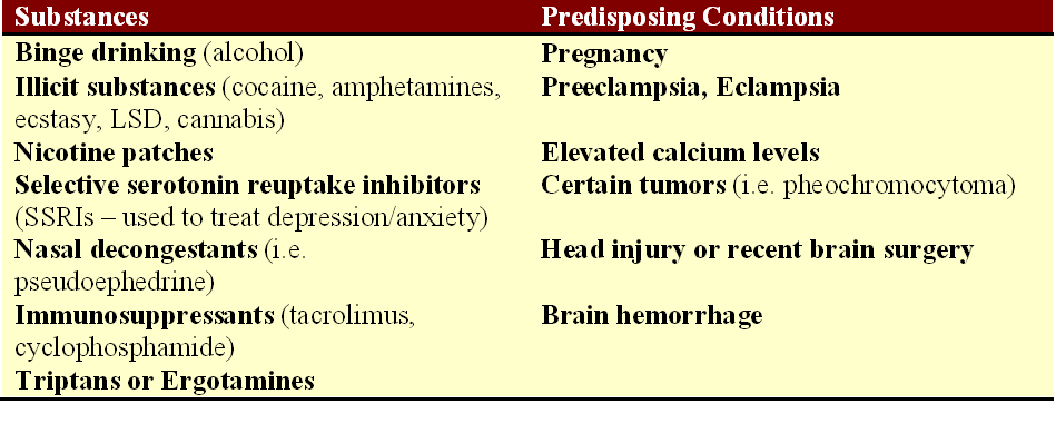
What causes RCVS? The exact cause of RCVS is unknown, but there are predisposing factors that can be associated with RCVS. Pregnancy and the postpartum state, particularly in women with preeclampsia, can be a trigger for the development of the condition. Selective serotonin reuptake inhibitors (SSRIs), frequently used to treat depression and/or anxiety, have also been associated with RCVS. The use of “vasoactive medications,” meaning medicines that can cause constriction of the arteries, can trigger this as well. This would include triptans, ergotamines (such as DHE for migraine), nasal decongestants that contain ephedrine or pseudoephedrine, certain immune suppression medications used in autoimmune disorders or after organ transplantation, or illicit substances such as cocaine, methamphetamine, ecstasy, and LSD. Cannabis has also been reported in association with thunderclap headaches resulting from RCVS.

RCVS triggers

Identified triggers for the development of reversible cerebrovascular vasoconstriction syndrome. Source: Tan and Flower. Emergency Medicine International, 2012.

Is RCVS a type of migraine? While approximately 40% of patients with RCVS report a history of migraines, thunderclap headaches that occur as part of the vascular constriction are not typical migraines. In fact, triptans that are typically effective in alleviating migraines can actually worsen the narrowing in the blood vessels that is occurring as part of the RCVS thunderclap headaches and should be avoided.

How is RCVS treated? For many patients, RCVS is a self-limited syndrome, and the headaches will stop after several weeks. However, some patients do experience recurrence or ongoing symptoms that may warrant intervention. The first thing that must be done is to remove the trigger, if known, for what may be causing the blood vessels to spasm. If a patient is taking an SSRI, it should be discontinued. Supportive care and pain management through the period of thunderclap headaches may be enough for some patients.  There are no randomized clinical trials to definitively answer the question of how best to treat RCVS, but calcium channel blockers (nimodipine, nicardipine, and verapamil are three such examples from this class) have been utilized with some success in the observational studies that are published. Magnesium may be helpful also, particularly in a pregnant or postpartum patient with eclampsia or preeclampsia.

I have seen this syndrome described as “rare,” but like so many syndromes that may result in stroke in younger patients, I ask myself – rare? Or underdiagnosed? I suspect the latter.

Understanding cerebral aneurysms following the death of journalist Lisa Colagrossi

A reporter for WABC in New York City, Lisa Colagrossi, died over the weekend after an aneurysm in her brain ruptured. Colagrossi, 49, apparently experienced the very sudden onset of symptoms, and was maintained on life support in the neurological intensive care unit at New York-Presbyterian Weill Cornell Medical Center. Unfortunately, despite the care provided, she did not survive.

meninges

Layers of the meninges covering the surface of the brain. It is in the subarachnoid space that the major arteries of the brain are positioned, and a ruptured aneurysm results in bleeding along the surface of the brain. Ref: http://www.merckmanuals.com

What happens when an aneurysm ruptures? Where does the bleeding take place within the brain?

The meninges, or the thin tissue coating the surface of the brain, have three basic layers: the dura (outermost layer, which is adjacent to the skull), the arachnoid, and then the pia (adjacent to the brain). When an aneurysm ruptures, typically it is into the subarachnoid space, meaning just beneath the arachnoid layer of the meningeal coating. Thus, a subarachnoid hemorrhage occurs when blood fills the space below the arachnoid layer.

Because the subarachnoid space covers the surfaces of the brain (not just the superficial surface adjacent to the skull, but the deeper surfaces positioned further from the skull as well), bleeding into this space tends to produce a characteristic pattern on a head CT scan that differs from bleeding that is confined the portions of the brain adjacent to the skull (click here for descriptions of other patterns of bleeding from a prior blog post). A head CT scan is a very useful tool in quickly evaluating a patient complaining of symptoms concerning for subarachnoid hemorrhage. The image to the right is a more severe example of this.

What are symptoms that are concerning for subarachnoid hemorrhage? Typically, I use the word “sudden” in describing what occurs. A sudden headache that goes from non-existent to “the worst headache of my life” within seconds, also known as a thunderclap headache, should be urgently evaluated as a subarachnoid hemorrhage (911 call, head CT scan, urgent work up) until it is proven to be otherwise. Sudden loss of consciousness can be consistent with this presentation. Sudden weakness or numbness on one side of the body, as might be seen in any type of stroke, can also reflect a subarachnoid hemorrhage in process. If a head CT scan is normal but the symptoms or history are still concerning for potential ruptured or “leaking” aneurysm, then a lumbar puncture is performed to evaluate for the presence of blood products (blood that is breaking down) in the patient’s spinal fluid. If concern persists, then a catheter cerebral angiogram, a test in which a catheter is threaded through the arteries, typically from the femoral artery in the groin, to image the arteries of the brain closely, may be performed.

SAH1

A head CT scan from a patient with a ruptured left middle cerebral artery aneurysm. Blood appears white on this form of imaging. Blood is layering along the various inner surfaces of the brain.

What causes aneurysms to form in the arteries of the brain? There are certain situations where genetics play a role, but the majority of ruptured aneurysms and subarachnoid hemorrhages occur in families without a history of similar occurrences. Modifiable risk factors, or activities that place people at risk that can be changed through behavioral changes, include cigarette smoking and chronic alcohol use. Hypertension has also been shown to increase the risk of aneurysm development. Smoking seems to be the universal risk factor, doesn’t it? But yes, cigarette smoking has been demonstrated as increasing the risk for aneurysm formation in the brain in numerous studies at this point.

What should be done if an aneurysm that has not ruptured is found? One of the challenges in evaluating patients with cerebral aneurysms is knowing when to pursue treatment of an aneurysm in the brain, and when to monitor the aneurysm. A common scenario is that of the patient experiencing headaches that turn out to be part of a migraine syndrome, but the headaches lead to imaging of the arteries of the brain. Then, a small aneurysm is identified. Now what? Evaluating this patient population involves weighing benefit against risk. If an aneurysm is quietly present, not causing problems, and the risk of rupture is extremely low, then the risk of a procedure to “fix” the aneurysm is not justified. If an aneurysm is at higher risk of rupture and hemorrhage, then the risk of intervening is outweighed by the benefit in securing the aneurysm and preventing a catastrophic hemorrhage. There are many factors to consider when making a decision about whether to intervene on an aneurysm. The location (the artery affected by the aneurysm) is important, as is size, the patient’s age, and the patient’s medical history.

Unfortunately, Ms. Colagrossi’s passing is another example of how anyone can be affected by stroke, and that a person is never too young to face this diagnosis. If her family and friends come across this at some point, I offer them my sincerest condolences on their loss.

Mistakes young stroke patients make

I will keep today’s post brief, but wanted to relay mistakes that young stroke patients frequently make in hopes that they will not perpetuate. Yesterday I saw a young stroke patient who decided to rest when symptoms began, so it is worth reiterating errors made and why these actions should be avoided.

  • Taking a nap/lying down when stroke symptoms begin. Remember, an ischemic stroke patient is only eligible for IV t-PA (the “clot busing” medicine) for 3-4.5 hours after a stroke begins, and with each passing minute that the brain does not receive blood flow, approximately two million cells will perish. When a stroke patient awakens from a nap, it is often too late to intervene. Call 911.
  • Driving himself/herself to the hospital. This is a terrible mistake for many reasons. A stroke patient is more likely to be involved in a motor vehicle collision if trying to drive while the brain is not receiving adequate blood flow/oxygen. Vision and cognition may be impaired. There could be delays in care with traffic on the road. Instead of being brought directly back to be evaluated by the emergency medicine physician, a patient may incorrectly be asked to wait in the triage area if not arriving by ambulance, which creates further delays. Do not drive to the hospital if you may be having a stroke.
  • Taking aspirin at home. Remember that 20% of all stroke are hemorrhagic, or “bleeding strokes.” Aspirin may worsen bleeding, and no one will know whether a stroke is ischemic (due to a blood clot blocking blood flow to the brain) or hemorrhagic until a head CT scan or MRI can be performed to visualize the brain.

I had the privilege of providing a basic overview about stroke on May 1, 2014 on a local news show in Charlotte, NC to start Stroke Awareness Month this year. If you are interested in viewing this, click here. Remember the FAST mnemonic for identifying stroke early so we can work to prevent disability from stroke.

  • Face – Is a facial droop present on one side?
  • Arm – If both arms are extended, and one drifts down due to weakness, this could be a stroke until proven otherwise
  • Speech – Is there slurred speech? Is there difficulty finding the words with which to communicate?
  • Time – Time is critical, as stated in the above points. Call 911.

A Normal Head CT Scan Does Not “Rule Out” Ischemic Stroke – Part II

As a follow up to last week’s post about head CT scans failing to demonstrate evidence of ischemic stroke in certain situations (early stroke, strokes of small sizes, strokes in the brainstem or cerebellum), I wanted to share several cases illustrating the truth behind the assertion.

CT negative stroke CThe head CT image on the right was obtained from a young woman who was 31 years old at the time of her stroke. She presented to an outside emergency department at a small hospital with numbness and jerking movements of her left arm. Her blood pressure was high, and she was discharged home with a diagnosis of hypertension. Her head CT scan was normal at that time. Shortly after arriving home, she developed prominent left-sided weakness, returned to the ER, and then was diagnosed with an early ischemic stroke. The patient’s right cerebral hemisphere (which is on the left side on our view – the patient is facing us on this CT image, so what we see as the left side is actually the patient’s right side) appears darker than in the left hemisphere. This is because edema (swelling) and ischemic injury have occurred in the brain. Several years later, she has reduced use of her left hand and struggles with anxiety. She and her husband have been a source of great inspiration to me as I have had the privilege of observing them persevere through her challenges. CT negative stroke D

The MRI to the left belongs to a woman who presented to an ER with isolated leg weakness, and a stroke was “ruled out” with a normal head CT scan. She returned home, and later returned when her symptom worsened. Her brain MRI demonstrates acute cerebral ischemia (injury emerging from lack of blood flow) in the territory of the right anterior cerebral artery (the white arrow tip indicates the location of the stroke). See my previous post, A Tale of Two Carotid Artery Dissections, and the Miracle of the Circle of Willis, for a tutorial on the major arteries bringing blood to the brain.

For all of the frustration that I hear from patients about strokes that have been missed on CT scans, though, I feel that it is necessary to celebrate when a not-so-obvious stroke is diagnosed early enough for what it actually is, and the patient is able to receive the appropriate therapy for the best shot at a positive outcome. It does actually happen, and I tell the following story to offer hope. Sometimes things run as they should on the front lines of medicine, and it is quite refreshing when things go right!

CT negative stroke BA woman suddenly became very dizzy and had difficulty speaking clearly. The alignment of her eyes became skewed, and she was unable to focus on a single target. Following this, she became confused and disoriented, and was unable to move her right side. Her symptoms were fluctuating, and those around her could not figure out in that moment exactly what was occurring. She was brought to a local hospital, and the ER physician, concerned for possible stroke, called the on call neurologist. The patient had the head CT scan to the right, which was interpreted as normal by the radiologist (and which I agree is unremarkable). The patient received IV t-PA immediately after the CT scan was able to exclude this as a hemorrhagic stroke, on the assumption that the patient was in the midst of an early ischemic stroke.

On the following morning, the patient’s right-sided weakness had completed resolved, her speech was clear, and her thinking and reasoning had returned to normal. The only symptom that remained were some visual abnormalities. At this point, that has improved as well.

ct negative stroke AHer brain MRI (on the left) that was performed the following day revealed that she had, indeed, sustained an ischemic stroke. Fortunately, she was able to receive IV t-PA early enough to minimize the damage. Thank goodness the emergency medicine physician did not “rule out” stroke with her normal head CT scan. On the following night, the same emergency medicine physician was working and called about a different neurological patient. During the discussion, feedback was provided to him about how the previous night’s patient had improved, and how the MRI did confirm the presence of ischemic stroke. His response was: “It always feels good to know that you’ve made the right decision.” Yes, it certainly does.

A Normal Head CT Scan Does Not “Rule Out” Ischemic Stroke – Part I

“Head CT ruled out stroke.”

It happens multiple times each week. A patient arrives for a consultation, bringing outside medical records from the emergency department where his or her initial evaluation took place. Inevitably I will find the above statement, or some slight variation of it, stated in the notes, as if serving as a justification for why it was okay to send the patient home. Often the patient wonders why he developed partial visual loss for 30 minutes, or why the left side of her body was tingling for hours even prior to the decision to seek medical attention. After all, the symptoms seemed real at the time, but how to explain what happened since the head CT ruled out a stroke?

From my own experience caring for young stroke patients, I hear the same themes reiterated:

  • “Everyone tells me that I don’t look like I’ve had a stroke, but I have never felt like the same person since my stroke.”
  • “I have so much difficulty focusing and concentrating since my stroke, and I’m worried about my short term memory.”
  • “I was told that I shouldn’t still be experiencing pain now that my vertebral artery dissection has healed, but my headaches are still awful.”

I could list at least 20 bulleted themes commonly heard when talking with young stroke patients, but perhaps the most common one that I hear is: “The first time I went to the ER, I was sent home.”

There are, of course, significant concerns about a young stroke patient seeking medical attention in the ER with stroke-like symptoms, and then being discharged home. The most critical reason to identify early ischemic stroke symptoms in a patient of any age is to take advantage of the only FDA-approved medication in the treatment of acute ischemic stroke – IV t-PA. As mentioned in a previous post, t-PA is a “clot-busting” medication. When a clot blocks blood from flowing in an artery, the administration of t-PA can dissolve the clot, relieving the obstruction and allowing blood to start flowing again. In acute stroke, every minute that passes equates to the death of approximately 1.5 to 2 million neurons (cells in the brain or spinal cord).

With each passing minute that blood is not flowing to part of the brain, the likelihood of a good functional recovery decreases. In addition to the decreasing benefit of t-PA with the passage of time during those first hours of an ischemic stroke, the risk of cerebral hemorrhage from giving t-PA increases. As tissue in the brain sustains injury from this lack of blood flow, the tissue becomes “leaky” and starts swelling, and giving a clot-busting medication like t-PA to a patient with brain tissue that has already died raises the risk that the drug could cause a brain hemorrhage.

ecass 3 graphThis graph appeared in the paper that outlined the results of a stroke clinical trial known as the ECASS 3 study. ECASS 3 was a trial that demonstrated that some patients benefitted in terms of functional recovery from receiving t-PA out to four and a half hours after the start of their stroke symptoms. Prior to this trial, the “window” for giving t-PA to an ischemic stroke patient was three hours after stroke symptoms began. The FDA in the United States has not approved t-PA for use beyond three hours into an ischemic stroke, although in other parts of the world t-PA has been approved for up to 4.5 hours based on this data. I include the graph from that paper to demonstrate that minutes do count. The higher the solid line is on the graph, the better the likelihood of a good functional outcome without significant disability after a stroke. As time passes on the x-axis (numbers on the x-axis represent the number of minutes that have passed since a stroke began), one can see the line trending down, indicating smaller chances of seeing a good outcome. Once the line crosses at 1.0, this is the time point at which is becomes more dangerous to give t-PA because the risk of bleeding is no longer outweighed by the benefit. At this point, for the majority of patients, brain tissue has died, and there is no more benefit to be gained.

When the assumption is made that a patient with stroke-like symptoms is not having a stroke, a CT scan of the head is usually obtained to confirm this assumption. This is flawed reasoning though. The sensitivity of a CT scan without contrast within the first 12-24 hours of an ischemic stroke is around 65%. This means that 35% of patients presenting 12-24 hours after a stroke has started will have essentially normal head CT scans. Now, consider patients who are within the time window for consideration for t-PA. The sensitivity of a head CT scan during the first three to four and a half hours of a stroke is even lower.

A head CT scan is a very good tool for diagnosing bleeding that occurs in the brain, and this is the reason why patients presenting with stroke symptoms are supposed to have a CT scan – to look for blood. The purpose of a CT scan should not be to diagnose an ischemic stroke. The likelihood of detecting hemorrhage in the brain is around 90-95% with a CT scan. Since 20% of strokes are of the hemorrhagic type, but it is impossible to determine based on a patient’s symptoms alone whether a stroke is ischemic (due to a blood clot blocking blood flow) or hemorrhagic, a CT scan clarifies the stroke type.

When I am seeing an acute stroke patient in the ER, I expect to see one of three things on the head CT:  1) evidence of bleeding (hemorrhagic stroke), 2) evidence of ischemic stroke (which means swelling and cell death has already occurred), or 3) a normal head CT scan. It is the patient with the normal head CT scan who should receive t-PA, because this is the patient who is very early into an ischemic stroke. An early stroke can be a treatable stroke, but if a patient is sent home on the assumption that a normal CT scan “ruled out” a stroke, then the only evidence-supported medical therapy for treatment has been denied to that person.