Young Stroke Survivors

Finally! A randomized clinical trial evaluating treatments for cerebral venous sinus thrombosis (CVST)

When the topic of stroke is being discussed, for the most part it is the concept of “blockages” developing within in the arteries of the brain – that is, the blood vessels that are carrying oxygen-rich blood *to* the brain to nurture its cells. These blockages can occur from the formation of blood clots, plaque build up, dissections (tearing of the lining of an artery’s wall and blocking blood flow), or a number of other rarer phenomena. Put simplistically, when these blockages occur, blood cannot supply a portion of the brain, and those brain cells, known as neurons, then die from the lack of oxygen and nutrients.

After blood cells have delivered oxygen to neurons, they then need to leave the brain to return to the heart, then travel from the heart to the lungs in order to pick up more oxygen, and thus repeat the cycle of oxygen delivery to the brain and other organs. Blood leaves the brain through drainage systems called cerebral venous sinuses (effectively large veins). Much less frequently than the development of blockages in the arterial system (about 2% of all strokes), blood can clot within these venous sinuses, resulting in what is known as a cerebral venous sinus thrombosis (CVST). When venous sinuses are blocked because of obstructions caused by blood clots within them, blood struggles to leave the brain and backs up, which can lead to brain swelling, bleeding, and in severe cases, coma and death. I discussed this topic in 2016 in the context of the US presidential election (click here if interested in reading more).

Venous sinuses within the brain drain blood and send it back to the heart. Image source: http://www.dartmouth.edu/~humananatomy/

The standard treatment for CVST has typically been to place patients on anti-clotting medications (known as “anticoagulation”), such as heparin, enoxaparin (Lovenox), warfarin (Coumadin), or more recently, physicians and healthcare providers are starting to use the newer anticoagulants such as dabigatran (Pradaxa), rivaroxaban (Xarelto), or apixaban (Eliquis). Most patients with CVST present to doctors or healthcare providers awake and thinking coherently, but can commonly experience severe headaches, blurred vision, and nausea and/or vomiting from the pressure that is building up within their heads as blood backs up and cannot drain. However, some patients come to medical attention with much more severe presentations, such as with difficult-to-treat seizures, severe confusion, or in comatose states. In this group of patients, at times doctors have gone with a more aggressive treatment approach, in which a wire catheter is inserted into the patient’s groin region and is threaded up to the site of the clot within a venous sinus of the brain to try to physically remove the clot. This is called a thrombectomy (“thrombus” refers to clot and “-ectomy” refers to the break up and removal). In some cases, t-PA, the “clot-busting” drug that can be used for arterial strokes, is infused at the actual site of the clot through the tip of the catheter in an effort to help dissolve a portion of the clot as part of the effort to physically remove it. Thrombectomy procedures have loads of data to support their use in carefully selected eligible patients who have obstructions in certain arteries of the brain, but had not previously been studied in a randomized clinical trial format in patients with severe CVST presentations.

Because CVST with severe clinical presentations are relatively rare, they have been traditionally difficult to study in order to determine whether these patients have better outcomes with only anticoagulation or with anticoagulation plus a more invasive procedure (thrombectomy and/or t-PA being infused into the clot, as described above). However, there is now a published clinical trial to offer some guidance in this scenario.

The TO-ACT trial was performed at eight hospitals across three countries, and was able to enroll 67 such patients presenting with CVST and severe clinical symptoms/findings. The primary outcome (the “final result” to see if the more aggressive treatment was a success compared to standard therapy) was to evaluate the number of patients at the end of 12 months who were normal neurologically or very close to normal, meaning they were fully independent and getting on with life. Thirty-three patients received anticoagulation plus they underwent thrombectomy, t-PA infusion, or both, while 34 patients only received only anticoagulation (no thrombectomy or t-PA). At 12 months, there was no difference in outcomes between the two groups. Going into the trial, the severity of symptoms/presentation was the same between the two groups of patients. Four patients died in the thrombectomy group, and one died in the anticoagulation only group, but the difference was not statistically significant (meaning, this difference could be due to chance, since the sample size was relatively small).

The authors of the study acknowledge that perhaps another larger study should follow. Let’s face facts, though – it is really difficult to enroll patients in a trial that is studying a relatively uncommon phenomenon. I personally find it impressive that this feat was able to be accomplished, and respect the persistence and determination among the investigators to bring this study to the finish line so that some guidance and insight could now be available into how to manage these patients. I have personally seen patients who show up in comatose states, looking very neurologically ill with CVST, who have done extremely well on anticoagulation, compared to how they appeared when diagnosed. I have seen others with “mild” presentations who have suffered with chronic headaches and other negative quality-of-life aspects that can really drag them down as they juggle life’s daily demands.

My bottom line with CVST is that earlier diagnosis is better. Once the diagnosis is made and patients are started on anticoagulation, the majority of people will go on to lead independent lives with their autonomy intact. It is a condition, unfortunately, that is frequently misdiagnosed, and it is tragic when the diagnosis is later made after seizures, coma, brain hemorrhage, or death occurs. Early diagnosis and treatment is perhaps the most critical factor in achieving good outcomes.

Stroke emerging as complication of Covid-19

On April 28, 2020, a letter published in the New England Journal of Medicine describing blood clots in large arteries supplying the brain (known as “large-vessel strokes”) in young patients testing positive for Covid-19 brought media attention to this particular facet of a disease physicians, healthcare providers, and researchers are endeavoring to understand. (Click here to read the brief NEJM letter.) The letter’s authors have been treating patients with Covid-19 in the Mount Sinai Health System in New York City. They note that, on average, every two weeks their hospitals typically encounter 0.73 patients with large-vessel strokes who are under the age of 50, but within a two week period, they encountered five, and all were Covid-19 positive. It’s a small sample size, but the trend is concerning.

As time passes and more information is published about what this disease looks like in its various presentations, it seems to start in many patients as a respiratory illness, but when patients become ill with it, it is because it has transitioned into a disease involving blood clots and/or severe inflammation within the arteries. Report after report has emerged of patients who are awake and talking with emergency medicine physicians and healthcare providers, who then quite suddenly go into cardiac arrest and die. Stories of patients losing limbs and fingers/toes due to blood clots are emerging. Lungs are full of blood clots (pulmonary emboli) in many patients. Stroke is yet another complication of blood that easily and readily clots when the brain is the organ on the receiving end of that impact.

And now children, who were initially thought to be safe from Covid-19, with “mild” or no symptoms initially, are being reported as developing inflammation within their arteries and going into heart failure, in what is similar to a syndrome known as Kawasaki disease.

(Click here for a good mainstream media article summarizing various effects of Covid-19 in layman’s language.)

As for my own patients – what I am personally hearing from people who have experienced strokes in the past who have been infected more recently with Covid-19 is that they seem to be losing the progress they made previously through rehabilitation and time. Old stroke symptoms from their prior brain injuries are rearing their ugly heads again, and deficits that they thought were in their rearview mirrors have now arisen again. This can happen after a stroke with any bodily infection, whether it is a urinary tract infection or pneumonia, and is called recrudescence.

As businesses begin the reopening process, we are still seeing tens of thousands of new cases emerging daily in the United States, and the death count in this nation now exceeds 78,000. Mind you, this is the death count. We’re not talking about patients who survive Covid-19 with long-term scarring in their lungs, or those who have suffered limb amputations or disability associated with strokes. We also still do not know what the long-term toll of this disease is. It was only about seven months ago that this virus began infecting human beings, and we really don’t know what the long-term effects will look like a year, five years, or ten years down the road.

I’m not trying to be unnecessarily alarmist or cause panic. I care about our economy. I deeply care about jobs, livelihoods, and businesses. But as a physician, I am being real.

I also know that people who need to get back to work will not be successful in the work place long-term if they are trying to recover from the aftermath of strokes, or if they have long-term breathing problems because of the lung damage they have sustained. I am looking at the Covid-19 pandemic as a long-term battle, and do not want all of the sacrifices that have been made to fight it to be in vain if/when it just keeps returning.

I mentioned in my previous blog post that Dr. Anthony Fauci announced to the press in March that models were projecting deaths of 100,000 to 240,000 people in the US, and reporters argued with him in disbelief. They didn’t like it. They didn’t accept it. He basically said he hoped for a better outcome, but the numbers were what they were. We are getting closer to this estimated range each day.

We can feel emotionally impassioned, bored, scared, or beyond ready to resume life as we knew it only a few months ago.

But unfortunately, viruses don’t play by our rules.

Fear accompanies loss of control, in Covid-19 and in stroke

In 2012, I walked through a cemetery in Cashion, Oklahoma, eager to find the final resting places of my grandmother’s seven siblings and many other long since departed ancestors and relatives. My grandmother, Alta Jane Anderson, was still alive then, going strong in her assisted living facility in Houston, Texas at the age of 92, her mind as sharp as it had always been. She could talk March Madness brackets each year, still read a book every week or two, and never forgot the birthday of any of her children or their spouses, her seven grandchildren or their spouses, or of her 18 great-grandchildren.

When she suffered her ultimately fatal hemorrhagic stroke at 96 years old on my son’s third birthday, April 21, 2016, my aunt put her on the phone with me, and she said, clearly in pain and her speech heavily slurred, “Hi, Sweetie. Is Alexander having a good birthday?” How in the world did she have the mental clarity or compassion in that moment to ask me that, instead of being concerned for herself? To this day, she carries the distinction of being the person containing the most genuine goodness within her of anyone I have ever known.

My final visit with my grandmother, 2016

As I walked through this sleepy rural graveyard, I called Granny on my cell phone, unsure of where to begin in my search for departed family members. Her voice lit up at the sound of mine, as it always did when any of her family called, and then she verbally navigated me to the correct area to pay my respects. Granny was born in 1919, and as I saw the grave stone of her older sister, Lottie, and the year of her death (1918, at age 20), the reality of my grandmother having never met her older sister enclosed around me. Lottie’s three month old infant son was buried next to her, having perished only four days before his mother.

I asked my grandmother what took both of their lives at such young ages. Her reply sounded like something out of a dusty history textbook, from a time in the very remote past: “It was the Spanish flu.”

(Interestingly, this pandemic received its nickname because the Spanish press was free to cover the impact of this flu, not because it originated in Spain.)

After finding other family grave sites, I wandered over to a shaded area beneath the canopy of a beautiful tree, and Granny and I proceeded to have a conversation about the great influenza pandemic of 1918. She recounted her mother’s stories of how her family could not have a proper funeral for Lottie and her son because of the fear around group gatherings, and the fear others had of catching this deadly flu. Burials were significantly delayed because of the high number of deaths within a short period of time, and local undertakers became overwhelmed.

As I sat in the cemetery on that gorgeous Oklahoma spring day, I distinctly remember having this thought: Thank goodness we live in the age of modern medicine and don’t have to live through the heartbreak of such a pandemic.

After all – the influenza pandemic of 1918 occurred during a time when only black and white photos were taken, almost a decade before the first “talking” movie premiered. We had advanced so far past that era.

As I write these words today on April 15, 2020, 33 days after schools shut down in Durham, North Carolina and group gatherings suspended, stemming from the same fears of more than a century ago, they seem naive. Even as a physician at that time, I was operating under the assumption that modern medicine could protect us from another pandemic. I knew that clinical trials and the development of vaccines take years to complete, but at that time, it seemed even to me that our technology, medicine, and people could spare us from another such pandemic of large magnitude.

And I still certainly hope that is the case. That historic pandemic took the lives of at least 50 million people worldwide, 675,000 of which were in the United States when its population was smaller than it is today.

Yet, despite our modern medicine, and brave, highly educated, and experienced doctors and healthcare providers on the front lines delivering care, the mortality rate as of the latest reported data in the United States from Covid-19 complications is at 4.2% (26,977 deaths and 617,628 cases – likely an underestimate of cases but an overestimate of the percentage of infections who die, given the lack of testing in people with mild or absent symptoms).

There is a fear that accompanies the Covid-19 pandemic such that most people have not witnessed anything like it in their lifetimes. I have, however, witnessed it in mine, on the faces of patients for whom I have cared who have suffered strokes at relatively young ages. I have seen numerous individuals who unexpectedly, while going through life in their primes, are stricken with a disease they thought only possible in older or unhealthier people. In particular, when a specific cause for the stroke cannot be identified, many of them initially live in daily fear, wondering if and when it will happen again.

Their fear stems from the unknown, and from the lack of control they have in their situations. Many of these healthy young people have asked me, “How is my cholesterol doing?” When I would inform them that their cholesterol had nothing to do with their strokes, often the question that would follow would be something to the effect of: “So what can I do not to have another one then?”

When Covid-19 existed primarily in China, much of the chatter in the western world among younger and healthier people involved words to reassure themselves that they would be safe, such as: “It’s really only older people and people with weak immune systems who die from this.” While older and immunocompromised patients are in the higher-risk categories for perishing from Covid-19, it didn’t stop the alarm many felt when stories began emerging of healthy young people who had succumbed to the disease. The 30 year old school teacher in New Jersey. The 39 year old disc jockey in Florida. The 17 year old in California. Eventually we learned that up to half of patients ending up in intensive care were under age 65. This did not fit our current ideas about flu, pneumonia, or any other passing viral contagious diseases to which we have become accustomed as a society. How, then, to ease the fear and gain a sense of control again?

What I have witnessed in stroke care since becoming a vascular neurologist in 2010 has been very similar. How do we explain it when a healthy 25 year old dies unexpectedly and quickly from a stroke when she had her whole life ahead of her? Is there a way of ever feeling in control again after witnessing such a thing?

Recently, I observed Dr. Anthony Fauci, the now-familiar face of the National Institute of Allergic and Infectious Diseases, speaking at a press conference, explaining that models were projecting a “best case scenario” of 100,000 to 240,000 deaths in the United States from Covid-19 if social distancing continued. Members of the press immediately launched critically into him. What they seemed to be saying was…we don’t accept that. That’s not right. He kept looking back at them, reiterating that the numbers were suggesting this, that he hoped for a better outcome with continued social distancing, but this is what the numbers suggested at that time, and that he was merely the messenger of this news. They argued with him. They didn’t like it.

As I watched, I thought – so many people in this country have not faced death. They have not seen the fear on a patient’s face as he or she wonders if today is his or her final day. This is the first time in the lives of millions of people when they have truly had to confront the question of how much time they have remaining, and they, understandably, find this distressing.

I have written before on The Stroke Blog about how I try to live my life to the fullest each day, not knowing if each day will be my last, because I have seen so many young people who think they have decades in front of them, only to find themselves struck unexpectedly with disability or death. It led me to a telemedicine career in 2018 so that I could savor my days with my children while they are still young, so they can remember their mother if that last day does arrive earlier than expected. It has led me to reflect on why stroke-related deaths in “young” celebrities impact us emotionally. Two years ago, stroke survivor Brett Patterson shared his story of searching for answers as to why he had a brain hemorrhage in his 20’s. He would be the first to tell you that he lives each day now with gratitude and perspective since that time, understanding that each day counts.

Silver linings exist in each human tragedy, as difficult as they may be to find. In the case of the Covid-19 story, perhaps a silver lining is bringing perspective to millions about the fragility of life such that we can learn to appreciate our days here, and understand what young stroke survivors have already known about the importance of making each day count. It’s the understanding that we can and should follow the recommendations of public health officials to prevent the spread of the virus and save as many lives as possible, but to find joy in the everyday-ness of life. Knowing that the ordinary is extraordinary. Having a conversation with a neighbor. Dining in a restaurant. Taking kids to school. Birthday parties. Working. Life.

Tomorrow is not guaranteed to any of us, global pandemic or not.

Reflecting On Stroke Anniversaries

Usually I associate the familiar phrase “The days are long, but the years are short” with the raising of children. It’s astonishing how exhausting days are with the sleep-deprivation that accompanies caring for newborn babies, chasing toddlers, or correcting a sassy pre-teen, but then – BAM!! – they are grown. Where do the years go when the days can seem so long? And as difficult as raising young children can be, when looking back on the challenges, there is an almost surreal quality to it.

It occurred to me recently that my patients have expressed to me this same sentiment with the process of stroke recovery. The long days in the hospital, then the acute rehab process early on with exhausting therapy sessions, then outpatient therapy while trying to return to life as “normal” when a new normal has arisen, and the previous normal no longer exists…wow. These days can drag on and seem absolutely interminable.

But then, the one year anniversary after surviving a stroke eventually arrives, then the two year anniversary, and then – five years have passed. The stroke anniversary remains etched in minds and hearts, and carries its own special significance. When the anniversary passes each year, it can bring many emotions.

  • Fear – “Will it happen again?”
  • Grief – “I miss the person I used to be and the life I used to have.”
  • Discouragement – “My doctor said I will stop improving after a year – does this mean I won’t continue to get better?”
  • Encouragement – “Look how much progress I have made!”
  • Victory – “Five years later, I’m making it each day – stroke isn’t going to stop me.”

(That whole you-won’t-improve-after-a-year thing is ridiculous, by the way, particularly in younger patients. Every person is unique is his or her recovery, and I have seen young patients still showing improvement three years later.)

Having heard and witnessed so much devastation that arises from stroke, I began feeling confused over time as some patients began sharing gratitude for their strokes. Grateful? For a stroke? How could that be? And as I have listened, the reasons have been numerous:

  • “I never would have chosen to have a baby if I had not had a stroke – it forced me to re-examine what I wanted in life.”
  • “I appreciate the little things so much more now than I did before my stroke.”
  • “I met the love of my life at a stroke support group.”
  • “I was so stressed out at work before my stroke and was burning at both ends. The stroke forced me to slow down and re-focus.”
  • “Before my stroke my house had to be absolutely perfect. The other day I found my two year old son coloring on the living room floor, and I was so grateful for him that it didn’t bother me.” (I have to remember this one patient’s story on a weekly basis.)

Perhaps this is what I find so inspiring about stroke survivors – the resilience in these statements, the search for the silver linings, and the way they keep going after unexpected life-altering events.

The Stroke Blog has been relatively silent recently as I have attempted to find my own sense of balance. Writing has always been a passion, and unfortunately it took a back seat to many other competing obligations over the past year. In the new year, though, it has moved closer to the front, and for that I am immensely grateful. I will be updating you on many topics, and look forward to the ongoing journey.

Here’s to a new year!

The Diagnosis I Never Expected: A Young Stroke Survivor’s Story

August 5, 2017 was a typical night for my girlfriend and me: quiet dinner, just the two of us, followed by dessert at Amelie’s, a popular bakery in Charlotte, North Carolina. After arriving home, little did we know that the night was only getting started.

At around 1AM, Jessie awoke to my arm twitching. I told her I couldn’t move it, but we thought I had just slept on it wrong. A few minutes later, I realized I couldn’t move the entire left side of my body. This was coupled with a noticeable facial droop. Panic started to set in, and neither of us knew what to do. Luckily, I had family close by, and my older sister came right over. The next thing I remember, paramedics were by my side and I was being rushed to a hospital.

Brett is seen here during his hospitalization after his brain hemorrhage. He has only a patchy recollection of his days spent in the hospital.

The following few weeks were a blur. I’ve been able to piece the events back together with the help of family and friends. I spent six days in the ICU, another week in intermediate care, and was then discharged to a rehab facility to start intense physical and occupational therapy for my arm and leg. While I don’t remember much about my time in the hospital, I do remember one morning a nurse coming in and taking sixteen tubes of blood in order to try to get some answers. Some routine tests, and some sent off to the Mayo clinic for a more comprehensive analysis. Regardless, the tests all had one thing in common – they came back negative.

While I was hospitalized, countless doctors and nurses would enter the room and perform scans, MRIs, and other medical tests that I didn’t even know existed. The only consensus was that I had a right-sided intracerebral hemorrhage in the basal ganglia (structures deep within the brain, a common location for bleeding, although not in someone my age), which resulted in left-sided weakness. It looked like your classic brain hemorrhage caused by high blood pressure, or so I was told.

There was only one problem: I was a healthy 28-year-old man with no history of high blood pressure. My blood pressure was not even high the night of the stroke when the paramedics evaluated me.

Even after being transferred to the rehab hospital, more doctors came in, more tests were done, but no diagnosis was reached. The two weeks I spent at the rehab hospital were both challenging and eventful. In that 14-day time span, my nephew was born six weeks early and my grandmother was in and out of the hospital twice for her own medical reasons. My mom was a trooper, running around to three different hospitals and getting very little sleep. Despite this, I never spent a night alone in the hospital. My family, friends, and girlfriend provided me with more support than I could have ever expected.

Brett learned how to walk again with the assistance and encouragement of many physical therapists along the way.

After two weeks in the rehab hospital, I was healthy enough to return home and start my outpatient therapy. I had entered unable to walk independently, and I left walking with a cane. I was able move my arm a few inches side to side, but otherwise it was unable to perform any tasks. However, I knew the recovery process was just beginning, and I was told by many doctors that I could make a full recovery since I was young and healthy. Hanging on to that notion was reason enough to work hard and keep moving forward.

While I was still hospitalized, one of my neurologists highly recommended to my mom that we travel to Duke University to see Dr. Dodds in order to try and find more answers. Even though I was starting to accept the unknown etiology, I wanted to exhaust all options before throwing in the towel. Within days of arriving home, I told my mom to make the call to Duke. Little did I know it would be the best decision I’ve made in a long time.

Three weeks later, I made the trip to Durham with my mom, girlfriend, and brother and met Dr. Dodds. She looked at my images and noticed right away that not only did I have a hemorrhagic stroke, but I had also suffered an ischemic stroke as well. She felt there must be a unifying diagnosis to explain the presence of both hemorrhage and ischemic strokes occurring as part of the same event. We sat down for over an hour going through the weeks leading up to that eventful night. I had been sick a few weeks prior to the stroke, going to the doctor several times for headaches, fever, and a rash on the right side of my body. Dr. Dodds said she wanted to ponder everything for a couple of weeks, talk to some colleagues, and get back to us with more answers.

At 9AM the very next morning the phone rang and it was Dr. Dodds. She said it hit her very early in the morning, and she thought she knew what caused my stroke. “Brett, have you heard of varicella-zoster virus?” With little medical background, I told her I was not aware of it. She went on to explain it’s the shingles virus (which explained my rash), and she thought the virus may have gotten into my spinal fluid, causing the brain bleed and thus, causing my stroke. Varicella-zoster virus can infect the blood vessels of the brain, causing both brain hemorrhages and ischemic strokes. She said there were cases published in the medical literature of patients with this particular problem with MRIs that looked very similar to mine. The only real way to confirm her theory would be through a spinal tap.

Lo and behold, the spinal tap confirmed the unimaginable. The virus was present in my spinal fluid. Who would’ve thought? Shingles virus getting into my spinal fluid and causing a stroke? Of all of the possible diagnoses for a brain bleed, I felt very fortunate that at least mine was treatable. Unlucky to have experienced this in the first place, but fortunate. Since that time, I’ve been on two rounds of antiviral medication and will go in for my third spinal tap in a few weeks. The viral numbers came down on the second tap, and they were getting closer to the ‘negative’ range on that study.

Brett traveled with his family to New York City in December 2017, his first time flying since his stroke. The first travel experience after a stroke is often a milestone for young stroke patients.

After lots of PT and OT, I sit here today typing this story with both hands. I’m able to walk on my own, traveled to New York City last month with my family, and have started working with a therapist on getting back to jogging, playing golf, and hopefully returning to work eventually.

I share all this publicly for one very simple reason – DON’T STOP LOOKING FOR ANSWERS. It would’ve been easy for me to accept the unknown etiology and just move on with my life. However, I needed the answers, if not for myself, for my family, and everyone who spent countless hours by my side helping me through this difficult time.

Although my diagnosis is rare and may not be relevant to all, I encourage any stroke survivor without answers as to what caused his/her stroke to explore all options. Seek a second opinion. Ask if there are case studies published that might relate to your stroke. Don’t stop until you are satisfied. I found my answers, and with the right help and guidance, you might find yours as well.


New book for patients with carotid and vertebral artery dissection is now available

One of the most meaningful parts of my neurology residency training was learning how to treat patients with carotid and vertebral artery dissections. Not only was I fascinated with the concept that a young person could sneeze or cough and severely injure an important artery as a result of such a benign action, but I was surprised by how frequently we identified dissections, yet they were referred to as “rare.” They did not seem rare to me, but I figured I was biased, given the specialty I had chosen to pursue.

Following my vascular neurology fellowship completion, I then moved to Charlotte, North Carolina, and continued to frequently make this diagnosis in young, healthy individuals. I found that many of them were presenting to the emergency departments in the area with headaches and/or neck pain, and were diagnosed as having migraines or muscular spasms until I would recommend imaging of the arteries in the neck before sending them home. When imaging studies would reveal dissection of a carotid or vertebral artery, there was usually relief from both patients and healthcare providers in identifying a treatable cause for symptoms, and in knowing that we could lower the risk of stroke from that point with the appropriate management. As I saw more dissection patients in the outpatient clinic setting, I learned that many of them continued to suffer with pain, anxiety, migraines, insomnia, fatigue, and difficulty concentrating, to name a few concerns. I heard these concerns voiced from dissection patients who had suffered strokes, and from those who had no evidence of stroke on MRI. I also observed that even patients whose follow up imaging indicated that their arteries were now “healed” they still had lingering symptoms.

Over time, I found that I was having the same conversations and answering the same questions repeatedly when talking to dissection patients and their loved ones. By 2013, I thought: “Someone should compile the questions these patients have in book form and attempt to answer them.” There was no such book available, and it seemed very much worth writing, if the right person would make the time for it.

On January 1, 2016, about six months after I had joined the faculty at Duke University, Amanda Anderson, a speech-language pathologist in Charlotte, a friend, and herself one of my former carotid artery dissection patients, contacted me, telling me she wanted to work on a “project” to distract her from the daily unrelenting pain that had come to define her dissection aftermath. She had already published a workbook series for patients with language impairment (aphasia), and I knew she would be a great collaborator. I decided that since the “right person” had not made time to write the book for patients that I thought needed to be written, then I would have to be that person.

We decided early into the planning process to write much of the book in a question-and-answer format, and that we would make it as comprehensive as possible, but provide explanations that were easy to understand. We also wanted the book to illustrate that carotid and vertebral artery dissection patients are real people with real lives, and found plenty of brave patients from around the country (and even one outside of the US) who were willing to share their stories. We wanted many of the personal stories to be written by the patients themselves in order to provide their perspectives in their own voices.

This morning, after a year and a half of writing and revising, Carotid and Vertebral Artery Dissection: A Guide for Patients and Their Loved Ones was published!

It has been such a moving, unforgettable journey, and a regular reminder of why I love this patient group so much. Amanda’s relentless enthusiasm and her compassion for her fellow dissection survivors has sustained me during busy times when it was challenging to find time to write.

I sincerely hope that if you or a loved one has experienced a carotid or vertebral artery dissection, this book provides answers to your questions, and perhaps just as important, validation of your experiences.

When A Baby Has A Stroke: A Personal Story From the Executive Director of International Alliance for Pediatric Stroke

“Your baby has a brain abnormality.”

Those were the chilling words my husband and I heard when I was 29 weeks pregnant with our third child. We were told by the perinatologist that our unborn baby’s brain ventricles were enlarged and she would probably have hydrocephalus, a condition that results when spinal fluid cannot leave the brain and can lead to increased pressure within the skull. He couldn’t tell us much more than that. We prepared for the worst and hoped for the best over the rest of my pregnancy.

Our daughter, Michelle, was born just shy of 36 weeks, and the neurosurgeon was at the delivery to confirm that she did, indeed, have hydrocephalus. Three days later, when the neurosurgeon placed a shunt (a “pump”) in her brain to divert the flow of spinal fluid, he came to us with “good” news. Her hydrocephalus was a result of a brain hemorrhage that she had suffered sometime during my pregnancy. Apparently, a hemorrhagic stroke was a one-time “event,” which meant she didn’t have any other underlying major medical conditions.

Or so we thought.

At three months old, we and the team of doctors following Michelle noticed that she wasn’t using her right arm. The first red flag. Babies should not show a hand preference before one year of age. Michelle was diagnosed with right hemiplegia (weakness on one side), which we later learned was a type of cerebral palsy. Three months later she started weekly occupational and physical therapy, which we were able to continue for over ten years! Our lives consisted of juggling two older children with Michelle’s therapy appointments, a leg surgery, many doctor visits, MRIs, and multiple ankle-foot orthotics as she grew.

This graphic from the American Heart Association/American Stroke Association is part of a public awareness campaign to inform the public that a person is never too young to have a stroke.

We were fortunate that Michelle’s stroke was diagnosed early so she could start therapy at a young age. It was also a blessing that we lived in the Chicago area with an abundance of medical specialists to help Michelle reach her full potential. Through these specialists, I was able to meet other families who also had a child that had suffered a stroke and start a local support group. Knowing that we weren’t alone was a tremendous benefit for us as parents and it allowed the kids to meet others just like them. We were also able to have some of these medical specialists donate their time to come meet with our parents at our local meetings.

Unfortunately, sometimes good things come to an end. When Michelle was ten we moved to the Augusta, Georgia area. Even though I thought I had done my homework and assembled a team of medical specialists for Michelle, we discovered that medical philosophies vary from state to state. Access to specialists and hospitals is also limited in rural, less metropolitan areas. I wasn’t able to meet as many families as I had in Chicago, so support became an online endeavor. Two years later we moved to Charlotte, North Carolina, and again had to start fresh with new doctors and yet another philosophy about treating children experiencing the effects of a stroke. It was also quite a task to integrate Michelle’s educational needs in each of the new schools.

After moving twice in two years I gave up trying to create local support and decided it was time to create a global community with medically-vetted information and resources. That is how International Alliance for Pediatric Stroke was conceived. I have connected with so many families worldwide and have been able to work with pediatric neurologists and incredible advocacy leaders to improve awareness and education. What I have learned over the years is first, there are thousands of children impacted by stroke worldwide and families are eager to connect. Second, the resources and research for this population are lacking. Third, the diagnosis of stroke in babies and children tends to be delayed. Michelle’s “brain abnormality” being discovered before birth is not typical. Often, the diagnosis of stroke in babies is not diagnosed until months or even years after birth. That means these babies are missing rehabilitation opportunities during that valuable time early in life when their brains are rapidly developing.

Mary Kay and Michelle Ballasiotes promote advocacy and raise awareness of the challenges presented by stroke in early childhood.

The consequences for missing the signs of stroke in children can be even more devastating. Stroke is one of the top ten causes of death in children, and unfortunately, I have heard from parents who have shared their heart-wrenching stories of their children not surviving because the signs and symptoms were initially missed. One of my organization’s recent projects was partnering with the American Heart/American Stroke Association to create fact sheets for infant and childhood stroke. The more education and awareness we can provide on pediatric stroke, the better off these children will be.

Michelle is now 19 years old, and she just completed her first year of college. She drives, swims, was in the marching band, played soccer, took ballet, babysits, pet sits, has had multiple part-time jobs, and has been a public speaker for pediatric stroke since she was nine years old. We didn’t know what our baby’s outcome would be when we first heard those devastating words. We still don’t know what caused her stroke, which is the case with most perinatal strokes in children. It has been a learning process to navigate this unchartered path, but I have met incredible, strong families over the journey, and am hopeful for the future of all children impacted by stroke.

Allison Pataki shares young caregiver’s viewpoint after husband’s stroke at 30

Today, Allison Pataki published a moving New York Times blog post about her husband’s stroke at age 30. Pregnant with their first child at the time of the event, she describes continuing to work productively, preparing for a newborn, and instantaneously becoming her husband’s caregiver following this unexpected circumstance that life threw their way. Click here to read her tale.

#Redshoes4youngstroke: A Call to Action!

One of the central missions of The Stroke Blog since it went live in October 2014 has been to provide information to those who have survived a stroke or strokes that occurred at relatively young ages. The very first post, “Deconstructing the Mini-Stroke,” recognized that while there may be symptoms more-or-less universal to some stroke types regardless of a person’s age, that the young stroke population tends to struggle differently through what I call their stroke aftermath.

One problem in the young stroke population is that, while we have a drug (IV t-PA) that can help to minimize the long-term aftermath of ischemic stroke when administered within three to four-and-a-half hours of stroke onset, many young patients do not receive it. A number of my young patients have told me that when their symptoms began, they decided to take a nap or wait it out, either because stroke was not on their radar, or because even if it was, they believed stroke to be a disease of the elderly. Those who do take their symptoms seriously and seek emergent medical attention can be misdiagnosed, because healthcare providers may doubt that a stroke can occur at young ages. For hemorrhagic stroke, early medical attention can result in better outcomes for different reasons. Perhaps an aneurysm has ruptured requiring urgent surgical repair, or a hemorrhagic stroke patient requires emergent blood pressure control. When stroke symptoms develop, regardless of a person’s age, emergent medical attention should be sought. In the United States, this means calling 911 (not driving oneself to the hospital).

According to the Centers for Disease Control, in 2009, almost one-third of stroke hospital admissions in the U.S. were for patients under the age of 65. The Center for Health Statistics estimates that $15.5 billion was lost in productivity in the U.S. in 2008 as a result of stroke patients having to leave the workforce. When stroke strikes a young adult, it costs these individuals personally on many fronts, but it also takes its financial toll at a national level.

This is a public health problem. I want young people to know that when sudden paralysis develops in an arm or a leg, the right thing to do is to get to a hospital as quickly as possible in hopes that t-PA treatment may be a possibility. For severe strokes resulting in large artery occlusions, or “blockages,” we now have very compelling clinical trial data telling us that using a catheter to remove the blood clot is very beneficial in some patients, but only when the stroke is treated early. A delay of even a few hours may make the difference between being dependent on others for care, or returning to independence.

I challenge you to raise awareness about this problem. I challenge you to find a red pair of shoes in your closet, or purchase an inexpensive pair

Wearing my red shoes at the International Stroke Conference about 20 minutes before this blog post!

Wearing my red shoes at the International Stroke Conference about 20 minutes before this blog post! #redshoes4youngstroke

of red shoes, or if this is too much of a financial burden, to spray-paint an old pair of shoes red. Wear them proudly. The more they stand out, the better. If you are asked about them, use the opportunity to share with the questioner that a person is never too young to have a stroke. If you have a personal story to share, I challenge you to be bold enough to share it. If you are hesitant about sharing it, then communicate to others that stroke is not a disease only affecting the elderly.

Take a picture of your feet in these shoes, and post it to social media – to Facebook, Twitter, whatever. Include the hashtag #redshoes4youngstroke when you post it. Tag others in your posts whom you feel will care about this cause and participate. If you have the financial means to do so, consider making a donation to the American Stroke Association, National Stroke Association, Young Stroke, or another not-for-profit organization you feel has been supportive of the young stroke population. I will watch for interesting red shoe pictures with the #redshoes4youngstroke hashtag to come along, and will repost some of them with permission on The Stroke Blog.

Wearing my red shoes at the International Stroke Conference about 20 minutes before this blog post!

Moments before hitting “publish” for this post – hoping to bring more awareness around the plight of young stroke patients.

I’ll start. While purchasing a pair of boots for the winter online in January of this year, amazon.com recommended these shoes to me. I thought this was bizarre, as they looked nothing like the boots I had just purchased, but then it seemed almost fated. Somehow amazon.com knew me better than I knew myself, and realized that I would want these there’s-no-place-like-home shoes. Indeed I did, because I instantly decided to call them my “stroke awareness shoes.”

I wore them a few times earlier this month while caring for patients in the hospital to see how people would react, and I received multiple comments each day from patient family members, people in elevators, other parents when I picked my son up from basketball practice. I practiced giving my 20 second spiel about a person never being too young to have a stroke, and it resulted in a number of engaging conversations. Some people even said they would join me in wearing red shoes to raise stroke awareness!

I also want to thank the neurology residents at Duke University, who are not only fantastic physicians, but who have been my sounding board as I have contemplated this. They have been full of great ideas!

I am currently attending the American Heart Association/American Stroke Association’s International Stroke Conference, and am wearing my red shoes. I am encouraged at the response I have gotten over the course of the morning, and feel certain this can extend beyond those who care for stroke patients.

With greater awareness comes greater funding for research, greater compassion for the plight of a group of survivors, and greater understanding of an issue that exists in our society. Let’s wear our red shoes!

Clinical trial shows no difference in stroke prevention between antiplatelet agents and anticoagulation in carotid or vertebral artery dissection

Due to the number of readers of The Stroke Blog who have identified themselves as having experienced carotid or vertebral artery dissections with or without stroke, I believe a clinical trial from the United Kingdom is very much worth sharing here.

The Cervical Artery Dissection in Stroke Study (CADISS – Markus HS, et al) set out to answer a question that has existed for many decades in the world of stroke management. When a dissection (a tear in the innermost tissue layer of an artery) of a carotid artery or vertebral artery (arteries in the neck that bring blood to the brain), how is a stroke best prevented going forward? In patients who have already had a stroke at the time the dissection is diagnosed, the goal is certainly to prevent further strokes from occurring. If a patient has not already sustained a stroke, then sparing that person a permanent brain injury is the top priority. It has not been entirely clear how to achieve these goals though. Should a patient be treated with antiplatelet agents (medications impairing platelet function), such as aspirin, clopidogrel (Plavix), another antiplatelet agent, or some combination of these? Or should a patient be treated with anticoagulation, a drug that actively prevents clotting, such as warfarin (Coumadin) or heparin?

I have heard many arguments on all sides. Some neurologists say that antiplatelet therapy is just as effective as warfarin but carries a lower risk of hemorrhage. Others say that antiplatelet therapy is not aggressive enough and anticoagulation with warfarin or heparin (or both) should be used. Some say to start on antiplatelet therapy or anticoagulation and if symptoms get worse to switch to whatever therapy was not initially used. Others argue for placing a stent in the dissected artery. Until recently, there was no clinical trial actually comparing antiplatelet therapy to anticoagulation in patients with carotid artery or vertebral artery dissection, and the medicine selected for treatment was based entirely on anecdotes and the bias of the treating physician.

In the CADISS trial, patients presenting to one of the participating medical centers in the United Kingdom who were diagnosed with carotid or vertebral artery dissection (with or without stroke) believed to have occurred within the seven days prior to presentation were randomized. Half of the patients were started on antiplatelet therapy, and half were placed on anticoagulation. The duration of treatment was three months. The endpoint was to determine how many strokes or deaths occurred in each group. Whether or not the patient had already had a stroke before enrolling in the trial, the endpoint was to see, once antiplatelet therapy or anticoagulation was started, how many patients went on to have strokes despite that therapy.

The CADISS trial, published in Lancet Neurology in April 2015, did not find a statistically significant difference in stroke prevention in patients presenting with acute carotid or vertebral artery dissections when treated with either antiplatelet therapy or anticoagulation.

The CADISS trial, published in Lancet Neurology in April 2015, did not find a statistically significant difference in stroke prevention in patients presenting with acute carotid or vertebral artery dissections when treated with either antiplatelet therapy or anticoagulation.

Two hundred fifty patients were enrolled (118 carotid artery dissections and 132 vertebral artery dissections). Interestingly, 52 of these patients were not found to have carotid or vertebral artery dissections when their radiology studies were carefully reviewed as part of the study, despite initially receiving that diagnosis. Of the 198 patients remaining, there was no significant difference in strokes between the two groups. There was one episode of symptomatic bleeding in the anticoagulation group (subarachnoid hemorrhage, or bleeding in the brain that occurs when an artery in the brain ruptures). There were no deaths in either group. Of the 198 patients with radiological evidence of dissection confirmed, there were only four total strokes following initiation of the designated medical therapy in both groups combined.

The CADISS trial began as a feasibility study – a trial to see if it was even feasible to enroll enough patients with a condition not diagnosed with frequency in the emergency department. This phase of the study was statistically sound and convincing enough not to proceed with a larger trial.

A trial comparing stenting to medication alone has not been performed, and given the low number of strokes in patients on medical therapy in the CADISS trial, it is unlikely that a dissection stenting trial will be performed any time in the near future.

For more details about arteries bringing blood to the brain and about carotid and vertebral artery dissection, please refer to a prior post on The Stroke Blog by clicking here.

The take-away points from CADISS are as follows:

  1. The overwhelming majority of patients with carotid and vertebral artery dissection, if started on either antiplatelet therapy or anticoagulation soon after the dissection has occurred, will not go on to have a stroke while on therapy during the aftermath following the vascular injury.
  2. There is no significant difference in stroke prevention in patients with carotid and vertebral artery dissection between those using antiplatelet medications and those using anticoagulation.

The trial does not address the myriad symptoms many dissection patients notice lingering after the injury – migraines, neck pain, and anxiety, to name a few. One trial cannot address every possible issue associated with a medical condition. However, physicians who care for young stroke patients with this particular vascular injury should be celebrating the fact that we finally have evidence-based guidance for preventing stroke in these patients.

Cerebellar stroke – it’s about more than coordination and balance

The traditional teaching about the role of the cerebellum has typically been that it coordinates movements and “fine tunes” them. It provides balance when walking, and stability of a hand when reaching for a glass of water. When the cerebellum sustains an injury or is malfunctioning, then the result may be gait disturbance, falls, dizziness, or tremor.

The cerebellum is featured in red in this image. Image credit: www.brainhealthandgames.com

The cerebellum is featured in red in this image. Image credit: http://www.brainhealthandpuzzles.com

The ideas above are what I learned in high school biology, in anatomy, and in physiology. Even throughout my neurology residency training, I largely thought of the cerebellum as a structure that provided balance and fine tuned movement.

It has interested me during my time in clinical practice to witness the fallout from cerebellar stroke, particularly in the younger stroke population, because it is often far beyond balance and movement. Yes, the symptoms mentioned above are often present in some form when the cerebellar stroke occurs, perhaps along with a headache and/or nausea. However, the patients who struggle with recovery for months or years following a cerebellar stroke often complain of symptoms that do not fit with the traditional concepts of what the cerebellum is supposed to be doing.

Some of the complaints I have heard from numerous cerebellar stroke patients are as follows:

– Many struggle with the same cognitive symptoms that patients with strokes injuring the frontal or parietal lobes experience, such as difficulty with focus and multitasking, and because of this, they complain of difficulty with short term memory retention.

– Other cognitive symptoms may exist as well, such as feeling overstimulated, or having difficulty following a conversation in a group of people.

– Difficulty with language fluency (aphasia) has afflicted cerebellar stroke patients in my own experience, and their frustration after being denied disability benefits is palpable.

– Some cerebellar stroke patients express that they are unable to dream any longer, or that when they close their eyes to picture a scene – being at the beach on a breezy day, or running through a field of grass and flowers – they are unable to mentally visualize such a thing.

– Sometimes their significant others claim these patients have demonstrated changes in their moods or personalities, and that their relationships seem different since their strokes.

MR images of Jonathan Keleher's brain (A and B). The black diamond-shaped void in images A and B reveals Mr. Keleher's missing cerebellum. The images on the right demonstrate the presence of a cerebellum in the space in a normally developed brain. Photo credit: Massachusetts General Hospital, courtesy of Jeremy Schmahmann for use on NPR.org

MR images of Jonathan Keleher’s brain (A and B). The black diamond-shaped void in images A and B reveals Mr. Keleher’s missing cerebellum. The images on the right demonstrate the presence of a cerebellum in the space in a normally developed brain. Photo credit: Massachusetts General Hospital, courtesy of Jeremy Schmahmann for use on NPR.org

Last month, as I was driving home from work one evening, I heard this segment on National Public Radio’s All Things Considered, and I thought – yes! I have to share this on The Stroke Blog with readers! This piece summarizes the complexities of the cerebellum so well for the public, and I hope those of you who read this will take a few minutes to listen to the segment if cerebellar injury is of interest.

The piece features Jonathan Keleher, a 33 year old man who was born without a cerebellum. In the segment, it is explained that Mr. Keleher struggles with emotional complexity, language, and other cognitive tasks beyond imbalance and impaired motor skills. However, because he received intensive physical and speech therapy at a young age while lacking a diagnosis, he was able to demonstrate the wonder of neuronal plasticity – the ability to utilize other parts of the brain to accomplish tasks normally dependent on the cerebellum. He walks independently, and he works in an office environment. He lives independently.

We like to believe that each function is neatly packaged within a certain compartment of the brain. Patients often ask: “If my stroke was here [pointing to a specific part of the brain], then what problems should I expect to have?” While some structures in the brain correlate more or less with certain functions, it really is not that simple, as evidenced by the complexity of the cerebellum, and by what a young man who lacks one has been able to accomplish in its absence. The brain is a large community of cells, an interdependent network that makes us who we are, and which enables us to survive from one second to the next.

Update on November 14, 2017: 

When I published the above blog post on cerebellar stroke in 2015, I never dreamed that it would become the most frequently visited page on The Stroke Blog day after day. The comments readers have posted in response to it, and the emails I have received from patients and their loved ones, have underscored the need for more resources about cerebellar stroke. I have heard you, and am working currently to create such a resource beyond a blog post. Stay tuned.

I have also received many emails from patients who have been diagnosed as having vertebral artery dissections believed to have caused their cerebellar strokes. Until recently, there was no largely comprehensive resource for patients struggling through the aftermath of vertebral dissection, but a few months ago, my speech language pathologist colleague, Amanda Anderson, and I published a book for vertebral and carotid artery dissection survivors (click here for more information) and their families in hopes that it would provide badly-needed answers to lingering questions. If you are a vertebral artery dissection survivor, I sincerely hope you find the book useful, and that it at least somewhat helps to validate your “new normal.”

Cerebellar stroke can be more difficult to accurately diagnose because the symptoms frequently don’t scream “Stroke!” the way that weakness on one side of the body or a facial droop may. I have seen cerebellar stroke patients in the acute setting diagnosed with migraine, benign forms of vertigo, intoxication, and substance abuse. When diagnosed early, situations leading to cerebellar stroke can be successfully treated with better outcomes for patients. Awareness of cerebellar stroke in both the community and amongst medical providers is critical for earlier diagnosis and more optimal management.

Demystifying the Patent Foramen Ovale (PFO)

It occurs at least every other week in my own clinical experience. A patient has experienced an ischemic stroke, and after a workup that fails to show significant atherosclerosis (“hardening of the arteries,” “plaque build-up”) in the arteries leading to the part of the brain injured by the stroke, and without obvious risk factors that could have resulted in stroke, an echocardiogram identifies the presence of a patent foramen ovale (PFO). Often patients are told a PFO is a “hole in the heart,” allowing clots to reach the brain that would otherwise end up in the lungs as their final destination. I see in their notes: “Stroke caused by PFO.” Some patients arrive to the appointment, already having determined they want their PFOs closed, and others have already concluded just the opposite – no “heart surgery.” The majority feel lost and are seeking answers. What is a PFO, and what is the significance of it?

The purpose of this post is outlined in the title – to remove some of the mystery from the PFO, although its potential effects and clinical associations with it are, indeed, still mysterious.

FetalHeart

In the fetal heart prior to birth, blood enters the right atrium (A). Some of this blood circulates through an opening called the foramen ovale (yellow arrow in image), and into the left atrium (B), bypassing the lungs. When the foramen ovale fails to close, it becomes known as a patent foramen ovale, or PFO. Image source: http://vhlab.umn.edu

foramen ovale (“FO” – if you will) is a very normal part of a fetus’s heart. When we are fetuses, prior to taking our first breaths as our developing lungs remain collapsed, blood bypasses the lungs and receives oxygen from the placenta. As blood enters the right atrium of the heart, it can bypass the lungs by traveling through the foramen ovale, an opening connecting the right and left sides of the heart. After reaching the left side of the heart, blood can then proceed to travel through the left atrium, then the left ventricle, and finally exiting the heart through the aorta. Other features of fetal circulation exist to enable blood to more efficiently reach the placenta, such as the ductus arteriosis, which allows blood to stream from the pulmonary artery directly to the aorta rather than first having to visit the lungs.

A “FO” becomes a “PFO” (“patent” merely means “open”) when that opening between the right and left sides of the heart fails to close completely.

A PFO is not a congenital heart defect. This is an important piece of information for stroke patients found to have a PFO to understand. Many of them arrive at their appointment, believing there is something wrong with their hearts. It is a normal part of fetal circulation to have a foramen ovale.

PFOs are common. Population studies indicate that 20-25% of people have a PFO, and the overwhelming majority of them will never experience a stroke related to this opening between the right and left atria in the heart.  However, when younger people with cryptogenic ischemic stroke (stroke without an identifiable cause) are screened for PFOs, one is found in about 40% of patients being evaluated. This finding has been confirmed in multiple studies.

Screening for PFOs. Typically, a PFO is identified on an echocardiogram, which is an ultrasound study of the heart. Sometimes if there is significant blood flow through the PFO this can be identified on a standard echocardiogram. However, much of the time this is found after a bubble study, or agitated saline injection is performed. The patient has an IV in place in the antecubital region (between the arm and forearm, in the arm opposite the elbow). A small amount of air is mixed vigorously with saline to create numerous small bubbles and then this is injected through the patient’s IV. If a patient does not have a PFO, the bubbles will be visualized under ultrasound as entering the right atrium of the heart, but do not enter the left atrium because they have traveled to the lungs, which filter them. However, if a PFO is present, in many cases bubbles will be visualized entering the left atrium. Typically two injections are performed: one at rest, and one following the Valsalva maneuver, which involves bearing down/straining, as if lifting a heavy load or having a bowel movement. This maneuver increases return of blood to the right atrium, and a PFO that may not be significantly shunting blood to the left atrium at rest may suddenly enlarge when increased filling takes place in the right atrium, so the bubble test may be more dramatic.

There are limitations though. Breast tissue, obesity, and other factors may limit the viewing capabilities through the chest wall to detect bubbles in the left atrium. Sometimes, for no obvious reason, the study is just not of the quality needed to determine if a PFO is present. A transesophageal echocardiogram involves sedating a patient and inserting the ultrasound proble down the patient’s esophagus for a different view of the heart. This is thought to be a more sensitive study. The limitation to this test is that a sedated patient typically cannot be engaged in the procedure to voluntarily perform the Valsalva maneuver, so a PFO that is only a significant presence during such a maneuver may be missed.

TCD bubble study

This image compares three transcranial Doppler studies in patients with positive bubble studies for PFOs. The top image is from a patient with a PFO that is likely very small and which only allows a small degree of shunting from the right to left atrium. Each red vertical line represents a microbubble that has crossed to the left atrium and been detected in the brain during the test. The strip in the middle is from a patient with a moderate amount of shunting – more red lines/microemboli detected, but each one is still discernible from those around it. The bottom strip demonstrates the “shower effect.” The PFO is large and there is so much shunting that the microemboli are too numerous to count. Image source: http://www.revespcardiol.org

Transcranial Doppler is a study using ultrasound that measures blood flow within the major arteries of the brain. The same bubble study described above during an echocardiogram can be performed while blood flow is monitored in the middle cerebral arteries of the brain, and the detection of microemboli (small “blips” heard during monitoring) following the injection indicates that some form of passage from right-sided circulation to left-sided circulation is taking place. The limitation here is that pulmonary shunts, or vessels carrying blood directly from arterial to venous circulation within a lung, can also give a positive bubble test since transcranial Doppler does not involve the direct visualization of bubbles. However, signals are detected later than expected if the shunting is occurring in the lungs (more cardiac cycles following the injection than the timing of when signals from bubbles crossing through a PFO would be heard).

Transcranial Doppler is sensitive in detecting a PFO, and if consistent with this should be followed up with an echocardiogram, if not already performed.

If a PFO is detected, what then? This can be a challenging question to answer, in part because it is impossible to prove whether the PFO actually played a role in the stroke, or if another factor was at play and the PFO is just receiving the blame unnecessarily.

The question of whether to continue a stroke patient with a PFO and without another obvious cause of stroke on medical therapy or to close the PFO has been tested in at least two randomized clinical trials – CLOSURE I and RESPECT.

The CLOSURE I trial failed to show a significant benefit to PFO closure over medical therapy. However, CLOSURE I enrolled patients with either evidence of stroke on brain imaging or patients with “TIA.” The reason I place the term TIA in quotes is because more than half of the consults I see for diagnosis of “TIA” do not turn out to be actual TIAs, but migraines, seizures, or other conditions that mimick TIAs. The point here is that many neurologists argued that the validity of CLOSURE I results was negatively impacted by potentially enrolling patients into the trial with PFOs who had not actually had a vascular event, and that this may have skewed the data.

RESPECT NEJM

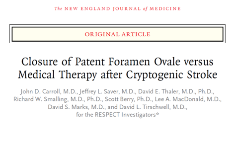
The RESPECT trial randomized young patients with PFOs and who sustained strokes without a known origin to either receive “best medical therapy” or to undergo PFO closure in addition to remaining on “best medical therapy.” There was no significant difference in stroke outcomes between the two groups, but may have been some benefit in patients with large PFOs or with aneurysmal atrial septum.

RESPECT was a trial randomizing only relatively young stroke patients with PFOs (no TIAs were included – there had to be evidence of stroke on the patient’s neuroimaging) who lacked another apparent source for the stroke to either PFO closure plus medical therapy, or medical therapy alone for stroke prevention. The overall trial failed to show benefit to PFO closure over medical therapy alone. However, in a secondary analysis, patients who had large PFOs or who had a finding known as an atrial septal aneurysm (very mobile wall separating the two halves of the heart) did show more potential benefit for stroke prevention from PFO closure and medical therapy than from medical therapy alone.

What is medical therapy? Medical therapy refers to any non-invasive therapy measures taken, in this case – to reduce the risk of another stroke. For patients with small PFOs and no history of stroke, this usually meant aspirin daily. For patients with atrial septal aneurysm, there is some data suggesting a relatively high risk of stroke recurrence on aspirin, but that anticoagulation may be more beneficial. I have found that the type of “best medical therapy” recommended to a patient often plays a role in his or her decision regarding PFO closure. Patients are often more willing to take aspirin daily over undergoing PFO closure, but many young patients are not thrilled with the idea of anticoagulation and the risks it carries over the long term.

It is mostly performed now via catheterization with an implanted device sealing the opening between the right and left atria. The procedure typically lasts less than one hour from start to finish, and patients usually go home from the hospital the following day. At some hospitals, they may even return home on the same day. There are potential complications to a PFO closure, including bleeding, infection, and the potential for atrial fibrillation, an irregular cardiac rhythm that can generate clots and result in stroke. Based on data from RESPECT, the incidence of atrial fibrillation did not differ significantly between the PFO closure group and the medical therapy alone group.

Following the publication of RESPECT trial results, PFO closure remains a topic of controversy in the field of stroke prevention and is still heavily debated.