Stroke Patient Tales

Reflecting On Stroke Anniversaries

Usually I associate the familiar phrase “The days are long, but the years are short” with the raising of children. It’s astonishing how exhausting days are with the sleep-deprivation that accompanies caring for newborn babies, chasing toddlers, or correcting a sassy pre-teen, but then – BAM!! – they are grown. Where do the years go when the days can seem so long? And as difficult as raising young children can be, when looking back on the challenges, there is an almost surreal quality to it.

It occurred to me recently that my patients have expressed to me this same sentiment with the process of stroke recovery. The long days in the hospital, then the acute rehab process early on with exhausting therapy sessions, then outpatient therapy while trying to return to life as “normal” when a new normal has arisen, and the previous normal no longer exists…wow. These days can drag on and seem absolutely interminable.

But then, the one year anniversary after surviving a stroke eventually arrives, then the two year anniversary, and then – five years have passed. The stroke anniversary remains etched in minds and hearts, and carries its own special significance. When the anniversary passes each year, it can bring many emotions.

  • Fear – “Will it happen again?”
  • Grief – “I miss the person I used to be and the life I used to have.”
  • Discouragement – “My doctor said I will stop improving after a year – does this mean I won’t continue to get better?”
  • Encouragement – “Look how much progress I have made!”
  • Victory – “Five years later, I’m making it each day – stroke isn’t going to stop me.”

(That whole you-won’t-improve-after-a-year thing is ridiculous, by the way, particularly in younger patients. Every person is unique is his or her recovery, and I have seen young patients still showing improvement three years later.)

Having heard and witnessed so much devastation that arises from stroke, I began feeling confused over time as some patients began sharing gratitude for their strokes. Grateful? For a stroke? How could that be? And as I have listened, the reasons have been numerous:

  • “I never would have chosen to have a baby if I had not had a stroke – it forced me to re-examine what I wanted in life.”
  • “I appreciate the little things so much more now than I did before my stroke.”
  • “I met the love of my life at a stroke support group.”
  • “I was so stressed out at work before my stroke and was burning at both ends. The stroke forced me to slow down and re-focus.”
  • “Before my stroke my house had to be absolutely perfect. The other day I found my two year old son coloring on the living room floor, and I was so grateful for him that it didn’t bother me.” (I have to remember this one patient’s story on a weekly basis.)

Perhaps this is what I find so inspiring about stroke survivors – the resilience in these statements, the search for the silver linings, and the way they keep going after unexpected life-altering events.

The Stroke Blog has been relatively silent recently as I have attempted to find my own sense of balance. Writing has always been a passion, and unfortunately it took a back seat to many other competing obligations over the past year. In the new year, though, it has moved closer to the front, and for that I am immensely grateful. I will be updating you on many topics, and look forward to the ongoing journey.

Here’s to a new year!

The Diagnosis I Never Expected: A Young Stroke Survivor’s Story

August 5, 2017 was a typical night for my girlfriend and me: quiet dinner, just the two of us, followed by dessert at Amelie’s, a popular bakery in Charlotte, North Carolina. After arriving home, little did we know that the night was only getting started.

At around 1AM, Jessie awoke to my arm twitching. I told her I couldn’t move it, but we thought I had just slept on it wrong. A few minutes later, I realized I couldn’t move the entire left side of my body. This was coupled with a noticeable facial droop. Panic started to set in, and neither of us knew what to do. Luckily, I had family close by, and my older sister came right over. The next thing I remember, paramedics were by my side and I was being rushed to a hospital.

Brett is seen here during his hospitalization after his brain hemorrhage. He has only a patchy recollection of his days spent in the hospital.

The following few weeks were a blur. I’ve been able to piece the events back together with the help of family and friends. I spent six days in the ICU, another week in intermediate care, and was then discharged to a rehab facility to start intense physical and occupational therapy for my arm and leg. While I don’t remember much about my time in the hospital, I do remember one morning a nurse coming in and taking sixteen tubes of blood in order to try to get some answers. Some routine tests, and some sent off to the Mayo clinic for a more comprehensive analysis. Regardless, the tests all had one thing in common – they came back negative.

While I was hospitalized, countless doctors and nurses would enter the room and perform scans, MRIs, and other medical tests that I didn’t even know existed. The only consensus was that I had a right-sided intracerebral hemorrhage in the basal ganglia (structures deep within the brain, a common location for bleeding, although not in someone my age), which resulted in left-sided weakness. It looked like your classic brain hemorrhage caused by high blood pressure, or so I was told.

There was only one problem: I was a healthy 28-year-old man with no history of high blood pressure. My blood pressure was not even high the night of the stroke when the paramedics evaluated me.

Even after being transferred to the rehab hospital, more doctors came in, more tests were done, but no diagnosis was reached. The two weeks I spent at the rehab hospital were both challenging and eventful. In that 14-day time span, my nephew was born six weeks early and my grandmother was in and out of the hospital twice for her own medical reasons. My mom was a trooper, running around to three different hospitals and getting very little sleep. Despite this, I never spent a night alone in the hospital. My family, friends, and girlfriend provided me with more support than I could have ever expected.

Brett learned how to walk again with the assistance and encouragement of many physical therapists along the way.

After two weeks in the rehab hospital, I was healthy enough to return home and start my outpatient therapy. I had entered unable to walk independently, and I left walking with a cane. I was able move my arm a few inches side to side, but otherwise it was unable to perform any tasks. However, I knew the recovery process was just beginning, and I was told by many doctors that I could make a full recovery since I was young and healthy. Hanging on to that notion was reason enough to work hard and keep moving forward.

While I was still hospitalized, one of my neurologists highly recommended to my mom that we travel to Duke University to see Dr. Dodds in order to try and find more answers. Even though I was starting to accept the unknown etiology, I wanted to exhaust all options before throwing in the towel. Within days of arriving home, I told my mom to make the call to Duke. Little did I know it would be the best decision I’ve made in a long time.

Three weeks later, I made the trip to Durham with my mom, girlfriend, and brother and met Dr. Dodds. She looked at my images and noticed right away that not only did I have a hemorrhagic stroke, but I had also suffered an ischemic stroke as well. She felt there must be a unifying diagnosis to explain the presence of both hemorrhage and ischemic strokes occurring as part of the same event. We sat down for over an hour going through the weeks leading up to that eventful night. I had been sick a few weeks prior to the stroke, going to the doctor several times for headaches, fever, and a rash on the right side of my body. Dr. Dodds said she wanted to ponder everything for a couple of weeks, talk to some colleagues, and get back to us with more answers.

At 9AM the very next morning the phone rang and it was Dr. Dodds. She said it hit her very early in the morning, and she thought she knew what caused my stroke. “Brett, have you heard of varicella-zoster virus?” With little medical background, I told her I was not aware of it. She went on to explain it’s the shingles virus (which explained my rash), and she thought the virus may have gotten into my spinal fluid, causing the brain bleed and thus, causing my stroke. Varicella-zoster virus can infect the blood vessels of the brain, causing both brain hemorrhages and ischemic strokes. She said there were cases published in the medical literature of patients with this particular problem with MRIs that looked very similar to mine. The only real way to confirm her theory would be through a spinal tap.

Lo and behold, the spinal tap confirmed the unimaginable. The virus was present in my spinal fluid. Who would’ve thought? Shingles virus getting into my spinal fluid and causing a stroke? Of all of the possible diagnoses for a brain bleed, I felt very fortunate that at least mine was treatable. Unlucky to have experienced this in the first place, but fortunate. Since that time, I’ve been on two rounds of antiviral medication and will go in for my third spinal tap in a few weeks. The viral numbers came down on the second tap, and they were getting closer to the ‘negative’ range on that study.

Brett traveled with his family to New York City in December 2017, his first time flying since his stroke. The first travel experience after a stroke is often a milestone for young stroke patients.

After lots of PT and OT, I sit here today typing this story with both hands. I’m able to walk on my own, traveled to New York City last month with my family, and have started working with a therapist on getting back to jogging, playing golf, and hopefully returning to work eventually.

I share all this publicly for one very simple reason – DON’T STOP LOOKING FOR ANSWERS. It would’ve been easy for me to accept the unknown etiology and just move on with my life. However, I needed the answers, if not for myself, for my family, and everyone who spent countless hours by my side helping me through this difficult time.

Although my diagnosis is rare and may not be relevant to all, I encourage any stroke survivor without answers as to what caused his/her stroke to explore all options. Seek a second opinion. Ask if there are case studies published that might relate to your stroke. Don’t stop until you are satisfied. I found my answers, and with the right help and guidance, you might find yours as well.


New book for patients with carotid and vertebral artery dissection is now available

One of the most meaningful parts of my neurology residency training was learning how to treat patients with carotid and vertebral artery dissections. Not only was I fascinated with the concept that a young person could sneeze or cough and severely injure an important artery as a result of such a benign action, but I was surprised by how frequently we identified dissections, yet they were referred to as “rare.” They did not seem rare to me, but I figured I was biased, given the specialty I had chosen to pursue.

Following my vascular neurology fellowship completion, I then moved to Charlotte, North Carolina, and continued to frequently make this diagnosis in young, healthy individuals. I found that many of them were presenting to the emergency departments in the area with headaches and/or neck pain, and were diagnosed as having migraines or muscular spasms until I would recommend imaging of the arteries in the neck before sending them home. When imaging studies would reveal dissection of a carotid or vertebral artery, there was usually relief from both patients and healthcare providers in identifying a treatable cause for symptoms, and in knowing that we could lower the risk of stroke from that point with the appropriate management. As I saw more dissection patients in the outpatient clinic setting, I learned that many of them continued to suffer with pain, anxiety, migraines, insomnia, fatigue, and difficulty concentrating, to name a few concerns. I heard these concerns voiced from dissection patients who had suffered strokes, and from those who had no evidence of stroke on MRI. I also observed that even patients whose follow up imaging indicated that their arteries were now “healed” they still had lingering symptoms.

Over time, I found that I was having the same conversations and answering the same questions repeatedly when talking to dissection patients and their loved ones. By 2013, I thought: “Someone should compile the questions these patients have in book form and attempt to answer them.” There was no such book available, and it seemed very much worth writing, if the right person would make the time for it.

On January 1, 2016, about six months after I had joined the faculty at Duke University, Amanda Anderson, a speech-language pathologist in Charlotte, a friend, and herself one of my former carotid artery dissection patients, contacted me, telling me she wanted to work on a “project” to distract her from the daily unrelenting pain that had come to define her dissection aftermath. She had already published a workbook series for patients with language impairment (aphasia), and I knew she would be a great collaborator. I decided that since the “right person” had not made time to write the book for patients that I thought needed to be written, then I would have to be that person.

We decided early into the planning process to write much of the book in a question-and-answer format, and that we would make it as comprehensive as possible, but provide explanations that were easy to understand. We also wanted the book to illustrate that carotid and vertebral artery dissection patients are real people with real lives, and found plenty of brave patients from around the country (and even one outside of the US) who were willing to share their stories. We wanted many of the personal stories to be written by the patients themselves in order to provide their perspectives in their own voices.

This morning, after a year and a half of writing and revising, Carotid and Vertebral Artery Dissection: A Guide for Patients and Their Loved Ones was published!

It has been such a moving, unforgettable journey, and a regular reminder of why I love this patient group so much. Amanda’s relentless enthusiasm and her compassion for her fellow dissection survivors has sustained me during busy times when it was challenging to find time to write.

I sincerely hope that if you or a loved one has experienced a carotid or vertebral artery dissection, this book provides answers to your questions, and perhaps just as important, validation of your experiences.

When A Baby Has A Stroke: A Personal Story From the Executive Director of International Alliance for Pediatric Stroke

“Your baby has a brain abnormality.”

Those were the chilling words my husband and I heard when I was 29 weeks pregnant with our third child. We were told by the perinatologist that our unborn baby’s brain ventricles were enlarged and she would probably have hydrocephalus, a condition that results when spinal fluid cannot leave the brain and can lead to increased pressure within the skull. He couldn’t tell us much more than that. We prepared for the worst and hoped for the best over the rest of my pregnancy.

Our daughter, Michelle, was born just shy of 36 weeks, and the neurosurgeon was at the delivery to confirm that she did, indeed, have hydrocephalus. Three days later, when the neurosurgeon placed a shunt (a “pump”) in her brain to divert the flow of spinal fluid, he came to us with “good” news. Her hydrocephalus was a result of a brain hemorrhage that she had suffered sometime during my pregnancy. Apparently, a hemorrhagic stroke was a one-time “event,” which meant she didn’t have any other underlying major medical conditions.

Or so we thought.

At three months old, we and the team of doctors following Michelle noticed that she wasn’t using her right arm. The first red flag. Babies should not show a hand preference before one year of age. Michelle was diagnosed with right hemiplegia (weakness on one side), which we later learned was a type of cerebral palsy. Three months later she started weekly occupational and physical therapy, which we were able to continue for over ten years! Our lives consisted of juggling two older children with Michelle’s therapy appointments, a leg surgery, many doctor visits, MRIs, and multiple ankle-foot orthotics as she grew.

This graphic from the American Heart Association/American Stroke Association is part of a public awareness campaign to inform the public that a person is never too young to have a stroke.

We were fortunate that Michelle’s stroke was diagnosed early so she could start therapy at a young age. It was also a blessing that we lived in the Chicago area with an abundance of medical specialists to help Michelle reach her full potential. Through these specialists, I was able to meet other families who also had a child that had suffered a stroke and start a local support group. Knowing that we weren’t alone was a tremendous benefit for us as parents and it allowed the kids to meet others just like them. We were also able to have some of these medical specialists donate their time to come meet with our parents at our local meetings.

Unfortunately, sometimes good things come to an end. When Michelle was ten we moved to the Augusta, Georgia area. Even though I thought I had done my homework and assembled a team of medical specialists for Michelle, we discovered that medical philosophies vary from state to state. Access to specialists and hospitals is also limited in rural, less metropolitan areas. I wasn’t able to meet as many families as I had in Chicago, so support became an online endeavor. Two years later we moved to Charlotte, North Carolina, and again had to start fresh with new doctors and yet another philosophy about treating children experiencing the effects of a stroke. It was also quite a task to integrate Michelle’s educational needs in each of the new schools.

After moving twice in two years I gave up trying to create local support and decided it was time to create a global community with medically-vetted information and resources. That is how International Alliance for Pediatric Stroke was conceived. I have connected with so many families worldwide and have been able to work with pediatric neurologists and incredible advocacy leaders to improve awareness and education. What I have learned over the years is first, there are thousands of children impacted by stroke worldwide and families are eager to connect. Second, the resources and research for this population are lacking. Third, the diagnosis of stroke in babies and children tends to be delayed. Michelle’s “brain abnormality” being discovered before birth is not typical. Often, the diagnosis of stroke in babies is not diagnosed until months or even years after birth. That means these babies are missing rehabilitation opportunities during that valuable time early in life when their brains are rapidly developing.

Mary Kay and Michelle Ballasiotes promote advocacy and raise awareness of the challenges presented by stroke in early childhood.

The consequences for missing the signs of stroke in children can be even more devastating. Stroke is one of the top ten causes of death in children, and unfortunately, I have heard from parents who have shared their heart-wrenching stories of their children not surviving because the signs and symptoms were initially missed. One of my organization’s recent projects was partnering with the American Heart/American Stroke Association to create fact sheets for infant and childhood stroke. The more education and awareness we can provide on pediatric stroke, the better off these children will be.

Michelle is now 19 years old, and she just completed her first year of college. She drives, swims, was in the marching band, played soccer, took ballet, babysits, pet sits, has had multiple part-time jobs, and has been a public speaker for pediatric stroke since she was nine years old. We didn’t know what our baby’s outcome would be when we first heard those devastating words. We still don’t know what caused her stroke, which is the case with most perinatal strokes in children. It has been a learning process to navigate this unchartered path, but I have met incredible, strong families over the journey, and am hopeful for the future of all children impacted by stroke.

Sports columnist Tom Sorensen returns after intracerebral hemorrhage

Tom Sorensen, a Charlotte-based sports columnist known for infusing wit into his writings on sports in the Carolinas, has returned to print after sustaining an intracerebral hemorrhage. He emerged back into print in The Charlotte Observer this week, opening up about his journey since tripping on a curb in September of this year.

His story is another example of why medical attention should be sought when something just does not seem right neurologically. It also may provide hope to those out there struggling to recover from a brain injury. Life can be good again.


Read Tom’s story by clicking here.

Can chiropractic manipulation cause stroke?

Since this story hit the news media last week, several blog readers have inquired into whether neck manipulation can result in stroke.

To summarize (and what I know is what the media is reporting – this is my disclaimer), Jeremy Youngblood, a 30 year old man in Oklahoma, died from complications of a cerebellar stroke following a visit to his chiropractor. While the media has not directly stated that Youngblood sustained a vertebral artery dissection, or a tear in the lining of the artery, this has been implied. The vertebral arteries (there are typically two – one on each side, traveling within openings through the vertebrae of the neck) supply blood to the brainstem and the cerebellum, which controls balance and coordination. Spinal fluid produced in the brain drains through a canal, called a ventricle, near the cerebellum. If a vertebral artery dissection occurs, inflammation occurs at that site in the blood vessel wall, and platelets begin to collect in that area. These platelet-rich clots then can break loose and travel to the brainstem and/or cerebellum, blocking blood flow to those sites, and resulting in ischemic stroke.

vertebral arteryThe danger in cerebellar stroke is that if swelling occurs in the area of damage and tissue expands, spinal fluid may not be able to leave the brain if the ventricle is closed off. The brain will still continue to produce spinal fluid, though, raising the pressure within the brain as a life-threatening condition called hydrocephalus occurs.

So – is there an increased risk of vertebral artery dissection following chiropractic neck manipulation? Many vascular neurologists would say they suspect the answer is probably, while acknowledging that many thousands of patients undergo this procedure without complications. In my own practice, I see two or three patients each year with a vertebral artery dissection that occurred somewhere in the midst of neck manipulation. However, often these patients sought help from their chiropractors in the first place for neck pain or headaches, so it is not possible to ascertain whether the dissection was already present. When patients state that within seconds of the manipulation stroke symptoms begin (sudden vertigo, nausea, vomiting, weakness on one side of the body, numbness on one side of the body, an inability to swallow – I have heard all of these as symptoms that have begun immediately following manipulation in patients later confirmed to have vertebral artery dissections with brainstem or cerebellar strokes on MRI), does that mean that the manipulation caused the dissection? Or was the dissection small, and worsened by the manipulation.

AHA scientific statement chiropractic manipulationRecently, the American Stroke Association issued a scientific statement about this controversial issue. In this statement, the claim is made that evidence is lacking to definitively associate the two, but that review of many population studies by the statement’s authors indicate an association between neck manipulation and vertebral artery dissection in young patients. The recommendation is that patients undergoing this treatment be informed of the potential, even if the risk is small.

Approximately half of vascular dissections are spontaneous – meaning, there is no explanation identified for how the lining of the artery, called the endothelium, was injured. In those where a source of trauma is identified, not infrequently the trauma is relatively minor. Some of the more interesting vertebral artery dissection stories I have heard are: head turning while swimming freestyle and feeling a “rip,” sneezing forcefully and experiencing sudden pain in the neck, lifting weights during a routine workout. There is the classic “beauty parlor dissection,” acquired when leaning back and extending the neck against the sink or dryer. One question is – how is it that a person can undergo the same activity 999 times without incident, and on that thousandth time be so unfortunate? What is it about circumstance and timing that makes the difference between a routine day at the gym (or at the chiropractor’s office), and the potential for stroke? While we know there are conditions that can place someone at additional risk for vascular injuries, such as Ehlers-Danlos syndrome or fibromuscular dysplasia, for the majority of vertebral dissection patients, there is no good explanation.

A Normal Head CT Scan Does Not “Rule Out” Ischemic Stroke – Part II

As a follow up to last week’s post about head CT scans failing to demonstrate evidence of ischemic stroke in certain situations (early stroke, strokes of small sizes, strokes in the brainstem or cerebellum), I wanted to share several cases illustrating the truth behind the assertion.

CT negative stroke CThe head CT image on the right was obtained from a young woman who was 31 years old at the time of her stroke. She presented to an outside emergency department at a small hospital with numbness and jerking movements of her left arm. Her blood pressure was high, and she was discharged home with a diagnosis of hypertension. Her head CT scan was normal at that time. Shortly after arriving home, she developed prominent left-sided weakness, returned to the ER, and then was diagnosed with an early ischemic stroke. The patient’s right cerebral hemisphere (which is on the left side on our view – the patient is facing us on this CT image, so what we see as the left side is actually the patient’s right side) appears darker than in the left hemisphere. This is because edema (swelling) and ischemic injury have occurred in the brain. Several years later, she has reduced use of her left hand and struggles with anxiety. She and her husband have been a source of great inspiration to me as I have had the privilege of observing them persevere through her challenges. CT negative stroke D

The MRI to the left belongs to a woman who presented to an ER with isolated leg weakness, and a stroke was “ruled out” with a normal head CT scan. She returned home, and later returned when her symptom worsened. Her brain MRI demonstrates acute cerebral ischemia (injury emerging from lack of blood flow) in the territory of the right anterior cerebral artery (the white arrow tip indicates the location of the stroke). See my previous post, A Tale of Two Carotid Artery Dissections, and the Miracle of the Circle of Willis, for a tutorial on the major arteries bringing blood to the brain.

For all of the frustration that I hear from patients about strokes that have been missed on CT scans, though, I feel that it is necessary to celebrate when a not-so-obvious stroke is diagnosed early enough for what it actually is, and the patient is able to receive the appropriate therapy for the best shot at a positive outcome. It does actually happen, and I tell the following story to offer hope. Sometimes things run as they should on the front lines of medicine, and it is quite refreshing when things go right!

CT negative stroke BA woman suddenly became very dizzy and had difficulty speaking clearly. The alignment of her eyes became skewed, and she was unable to focus on a single target. Following this, she became confused and disoriented, and was unable to move her right side. Her symptoms were fluctuating, and those around her could not figure out in that moment exactly what was occurring. She was brought to a local hospital, and the ER physician, concerned for possible stroke, called the on call neurologist. The patient had the head CT scan to the right, which was interpreted as normal by the radiologist (and which I agree is unremarkable). The patient received IV t-PA immediately after the CT scan was able to exclude this as a hemorrhagic stroke, on the assumption that the patient was in the midst of an early ischemic stroke.

On the following morning, the patient’s right-sided weakness had completed resolved, her speech was clear, and her thinking and reasoning had returned to normal. The only symptom that remained were some visual abnormalities. At this point, that has improved as well.

ct negative stroke AHer brain MRI (on the left) that was performed the following day revealed that she had, indeed, sustained an ischemic stroke. Fortunately, she was able to receive IV t-PA early enough to minimize the damage. Thank goodness the emergency medicine physician did not “rule out” stroke with her normal head CT scan. On the following night, the same emergency medicine physician was working and called about a different neurological patient. During the discussion, feedback was provided to him about how the previous night’s patient had improved, and how the MRI did confirm the presence of ischemic stroke. His response was: “It always feels good to know that you’ve made the right decision.” Yes, it certainly does.

A Tale of Two Carotid Artery Dissections, and the Miracle of the Circle of Willis

Nature is kind to some, and not so merciful to others. Much of what happens to us on a daily basis stems from chance, and almost every evening I try to reflect at least briefly on my good fortune to have experienced another day with my family, friends, and patients, knowing that it can all change so abruptly.

In order to understand the wonder and tragedy that lies within the following anecdote, it is necessary to understand some of the basic vascular anatomy of the brain – the plumbing that enables blood to circulate, that sustains the function and viability of the organ that makes us who we are.

circle of willis image

Image credit: http://www.deviantart.net

There are typically four arteries that bring blood to the brain – an internal carotid artery on each side (A) and a vertebral artery on each side (B); the vertebral arteries join together to form the basilar artery (C), which supplies blood to the brainstem and the back of the brain. The major arteries that extend from the internal carotid arteries in the brain are the middle cerebral arteries (D), and again, there is one on each side. The middle cerebral arteries supply blood to approximately 2/3 of the cerebral hemispheres. Smaller arteries supplying the inner midline surfaces of the brain also arise from the internal carotid arteries, called the anterior cerebral arteries (E). In the majority of people, there is a bridge connecting the right and left anterior cerebral arteries, called the anterior communicating artery (commonly called the AComm, F). With the AComm comes a connection between blood flow going to the right and left sides of the brain. I have seen patients arrive with completely blocked carotid arteries and absolutely no symptoms, and when evaluating cerebral blood flow with ultrasound or with angiographic imaging, it is clear that the side of the brain with the carotid artery occlusion is borrowing blood from the other side, using what is called collateral flow.  There is also the potential for sharing blood between the front of the brain and the back, via posterior communicating arteries (PComms, G, potentially one on each side). A complete circle in patients with both an AComm and PComms in addition to the other normal arteries in the brain is known as the Circle of Willis. About 30% of people possess a complete Circle of Willis.

During a weekend when I was on call, a previously healthy man in his 40s transferred from an outside hospital for management of an extensive right hemispheric ischemic stroke. He was critically ill upon arrival with an extremely concerning neurological exam. His angiographic imaging (imaging of his arteries) revealed that he had a right internal carotid artery dissection (essentially, tearing of the inner lining of the artery) with thrombus (clot) obstructing flow. His right hemisphere was ischemic, and he was unable to generate alternative ways of obtaining blood flow to this region in his brain. He lacked a complete Circle of Willis – no AComm and no PComms.

I find that certain days exist in which neurological themes declare themselves. On some days, I will see four patients consecutively who all have atrial fibrillation, an irregular heart rhythm that can result in ischemic strokes. On other days, I may receive three referrals for stroke that occurred during pregnancy or in the postpartum period. On that particular weekend, though, the theme was clearly carotid artery dissections. As I was leaving this man’s room in the neurological intensive care unit, the ER paged me, requesting guidance on a woman who had arrived with drooping of an eyelid (ptosis), and her pupil on the same side was more constricted than in the eye on the other side. This sounded like a classic Horner syndrome. Horner syndrome can occur with an injury to the internal carotid artery, which can result in both of the findings mentioned as well as abnormal sweating on the affected side of the face. I recommended that the patient have a brain MRI along with an MR-angiogram of the head and neck to exclude the possibility of a carotid artery dissection, a potential cause of Horner syndrome. I received a page soon after this that her imaging was, indeed, consistent with a significant carotid artery dissection. Fortunately, there was no stroke present. Aside from the eyelid droop and abnormally sized pupil, she was completely neurologically normal. Her MR-angiogram of the brain revealed that she possessed something valuable that the first patient lacked – she had an AComm.

MRA right carotid dissection

The blue arrow delineates flow in this patient’s normal left internal carotid artery. The green arrow demonstrates the absence of blood flow in the right internal carotid artery. The red arrow identifies the AComm, connecting the left (normal) circulation in the brain to the right side, and enabling the patient to avoid a stroke.

When I evaluated the patient in the ER, she told me that she became concerned about the abnormal appearance of her pupil, and had decided to seek medical attention for this in the ER rather than waiting until Monday to talk to her primary care provider. She was started on antiplatelet therapy for stroke prevention, and did not sustain a stroke. Nearly one year later, her Horner syndrome has improved (although not completely resolved) and she is doing well.

I am convinced that carotid artery dissections are underdiagnosed. Frequently, these patients present to the ER or to their primary care providers with headaches that seem like migraines at first glance. Perhaps a head CT scan is performed, which is not an effective study for excluding ischemic stroke (especially very early stroke). A brain MRI is a more sensitive tool for identifying early ischemic stroke, but if a carotid artery dissection is present, this will only be seen on a brain MRI if the dissection is located close to the brain and is large. If the dissection is small, or located farther down in the neck, a brain MRI will miss it. If a patient seeks medical attention for a severe headache unlike any other previously experienced, a migraine that differs from the patient’s description of his/her typical migraines if the person is already a migraine sufferer, or if the patient has any associated neurological symptoms involving the face, eyes, speech, or extremities, a brain MRI along with some sort of imaging of the arteries in the head and neck should be performed.

I reflect back on the ironic juxtaposition of these two patients presenting on the same morning, and marvel at what a difference the presence of a tiny artery in the brain can make. In this case, it was the difference between returning to a good, normal life without stroke, and tragically in the first patient’s case, an untimely death. He passed away within the week, despite the intensive medical care that he received.

I have often wondered if I have a complete Circle of Willis. Do I have an AComm? I don’t know. I do not have an answer, and perhaps I will never seek one, because what is up there is working for now, and I cannot change what I have. Certain cards are dealt to us long before we ever take our first breaths in the world, and as much as we like to feel empowered to create our own destinies, we do not have a say in whether our Circles are complete or not. Stroke brings the realization of a loss of control, and with this comes fear and anxiety. Stroke is more than just a diagnosis with symptoms. But when I see a patient with a carotid artery dissection and an Acomm providing flow, I see perseverance, and it gives me pause.

Deconstructing the “Mini-Stroke”

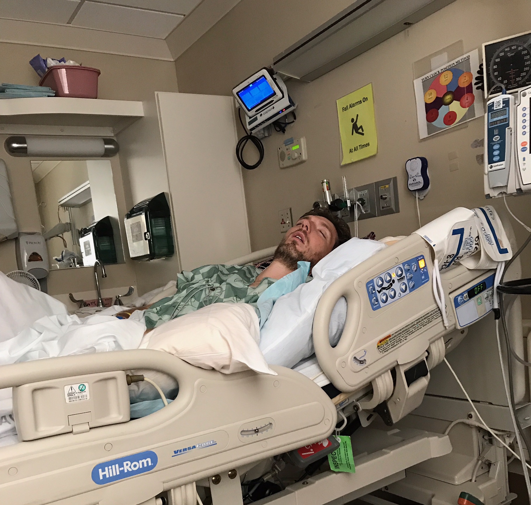
On Sunday, November 3, 2013, the Houston Texans played an NFL home game against the Indianapolis Colts, which began relatively uneventfully. However, it became clear that the game would not proceed in a typical manner as millions of viewers observed live footage of Texans head coach Gary Kubiak, 52, collapse on the field while exiting to the locker room at halftime.

The following headlines filled the national media during the week after Kubiak’s event:

TIA “mini-stroke” may increase risk for serious stroke – CBS News, November 6, 2013

Texans’ Kubiak Had Mini-Stroke, out Indefinitely – Associated Press, November 5, 2013

 Kubiak Released From Hospital After Suffering Mini-Stroke – 740AM KTRH Houston, November 5, 2013

Gary Kubiak Had Mini-Stroke: Houston Texans Coach Released From Hospital, Out Indefinitely – The Huffington Post, November 5, 2013

I am a vascular neurologist, a neurologist who has completed additional fellowship training in order to specialize in the treatment and prevention of stroke. Since completing my stroke fellowship in 2010, I have had the great privilege of caring for many patients just like Gary Kubiak, adults on the relatively young side who never expect such an event to disrupt their lives. The unfortunate reality is that stroke can happen at any age and can affect anyone.

A concern I felt with the headlines above is the reference to Kubiak’s event as a mini-stroke. This is a term that has become very popular in our American culture, and I hear it all of the time from my patients and their family members. “It was just a slight mini-stroke.” “Aunt Mildred had a mini-stroke while eating dinner.” “He had a touch of the mini-stroke.”

A stroke is a stroke. Period. A stroke results in an injury to the brain. There are two basic types of strokes – ischemic and hemorrhagic. An ischemic stroke occurs when blood cannot reach part of the brain for a prolonged period of time and permanent damage to brain tissue takes place. A hemorrhagic stroke occurs when a blood vessel ruptures and bleeding occurs within the brain. Roughly 80% of strokes are of the ischemic type.

A transient ischemic attack, or TIA, occurs when blood flow is disrupted to the brain and symptoms concerning for stroke occur, but then blood flow is either restored or the brain compensates for the absence of blood flow by seeking and acquiring blood from other sources and no damage to the brain occurs.

During a stroke, brain damage occurs. During a TIA, damage does not occur.

What about a TIA during which damage does occur? What is that called? The answer is – a stroke.

To describe a TIA as a “mini-stroke” misses the difference between the two terms. A TIA is not a stroke because damage is avoided. A stroke is not a TIA because brain damage has occurred. I like to refer to a TIA as an almost-stroke as opposed to a mini-stroke. Throughout the lifetime of this blog, I will continuously refer to TIAs as almost-strokes.

Sometimes patients may refer to a stroke with relatively mild deficits as a “mini-stroke” to distinguish it from a stroke that leaves someone externally and obviously disabled. This is also inaccurate. I have seen patients without a single physical visible deficit from a stroke who are significantly disabled from the cognitive impairment that frequently occurs following a brain injury. I have cared for a patient for the past two years whose only symptom from her “mini-stroke” (the term she used at her first appointment with me) was a left-sided neglect syndrome. This occurs when the brain fails to recognize that the left side of the body exists, even though the left arm and leg may move appropriately and strength on the left side can be left fully intact. She was a successfully employed person prior to her stroke in her 50s, and she has not been able to work since her stroke. She does not factor in columns on the left half of the screen when working with spreadsheets because her brain fails to recognize the left half of her conceptual world. She neglects to brush the left side of her hair and has tooth decay in the left side of her mouth because she does not brush her teeth on that side. She cannot drive because she visually neglects cars that appear in the left half of her world, even though her vision on the left side is intact. Is this really a mini-stroke?

In the initial evaluation of a stroke patient, this graphic is presented in order to calculate a score known as the National Institutes of Health Stroke Scale Score (NIHSS Score). The examiner asks the patient to describe what is seen in the picture as a test of language fluency. However, patients with profound left visual neglect will describe the woman washing dishes at the sink, but will fail to recognize the children in the left half of the scene.

In the initial evaluation of a stroke patient, this graphic is presented in order to calculate a score known as the National Institutes of Health Stroke Scale Score (NIHSS Score). The examiner asks the patient to describe what is seen in the picture as a test of language fluency. However, patients with profound left visual neglect will describe the woman washing dishes at the sink, but will fail to recognize the children in the left half of the scene.

Perhaps the other reason why I prefer to avoid the modifier “mini” in front of a word as significant as “stroke” is because patients tend to downplay the importance of the event. I love caring for patients after TIAs, because the damage has not yet occurred, and we can intervene to prevent a stroke! If a patient has a TIA and refers to it as “mini,” then I find there is less motivation for the person to quit smoking, comply with therapy, eat healthily, or exercise regularly. After all, it was only a mini-stroke.

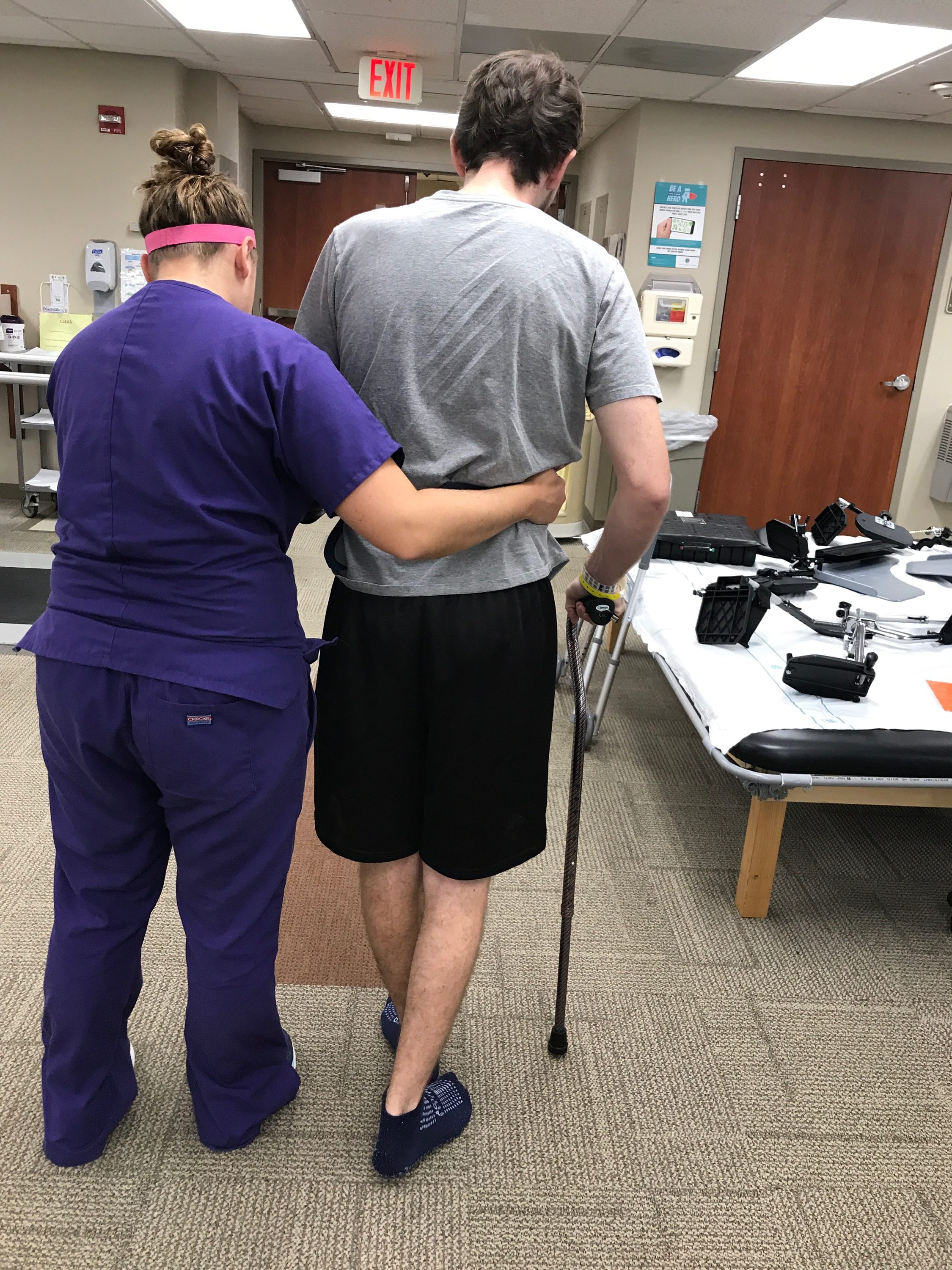
The other piece to these headlines is the relatively young age of the Texas coach. I definitely see patients at 52 with accelerated atherosclerosis (plaque buildup in the blood vessels, or “hardening of the arteries”), high blood pressure, diabetes, elevated cholesterol levels – some of the more typical stroke risk factors seen in older adults. However, it brings to light that a person is never too young to have a stroke, and more awareness hopefully will result in a call to 911 when stroke symptoms develop as opposed to taking a nap in an effort to sleep it off. Young people frequently do not believe their symptoms might represent a stroke, and choose to rest in hopes that the episode will resolve spontaneously. When they awaken, often there are no interventional options available, and therapy shifts from acute treatment of the current stroke to rehabilitating more long lasting deficits and focusing on how to prevent the next stroke.

During my stroke fellowship at the University of Washington/Harborview Medical Center in Seattle, I experienced my first encounter with stroke in the truly young patient. Samantha (not her actual name) was 16 years old when she developed weakness on the right side of her body and was diagnosed with an ischemic stroke based on her Magnetic Resonance Imaging (MRI) of her brain. Her physician recommended that she start taking aspirin daily, a medicine that assists in “preventing platelets from sticking to plaque in the blood vessel wall and from sticking to one another.” I put this phrase in quotes because I use it often when explaining why healthcare providers use aspirin for the prevention of strokes and heart attacks. While Samantha took her aspirin compliantly, she had a second stroke. Clopidogrel (trade name: Plavix) was added to her daily medication regimen. Take what I said aspirin does, and for most people, the effect is essentially more robust with clopidogrel.

Biological warfare had essentially been declared on Samantha’s platelets. She was also started on a statin, a class of medications to lower cholesterol and to protect blood vessels from accumulating plaque, or to protect against “hardening of the arteries.” Surely she was not going to have another stroke. But she did.

This is when I had the privilege of meeting Samantha. My stroke fellowship had just begun, and in my first weeks I encountered this young girl and her frightened mother. I did not know where to start, and I remembered words from mentors throughout medical school and my neurology residency training: Ninety percent of the relevant information for solving a medical mystery is in the history, or what the patient tells us. The rest of it – lab tests, radiology studies, you name it – confirms or denies a healthcare provider’s assessment of what the patient has described. Essentially, if the patient is describing symptoms of a stroke – weakness on one side of the body, drooping of the face, slurred speech – then my tests are to confirm or deny my suspicion. This is why taking the time to listen to patients is critical in providing care.

When Samantha recounted the stories of each stroke, there seemed to be a strong headache element. Headaches can occur with strokes, but many strokes are painless, and it seemed important that she would remember headaches while experiencing stroke symptoms. Not infrequently patients with strokes do not immediately recall a headache, because they are so alarmed by the weakness, numbness, or slurred speech that they forget to tell their physicians about the headache.

Samantha began her tale of her first stroke with details about a headache. My fellowship mentor and I decided that her strokes probably did not stem from platelets, blood clots, or plaque accumulation in her blood vessels. She was 16 years old, so how much plaque could she possibly have accumulated at that point in her young life? We determined that her strokes may have resulted from blood vessels constricting, or spasm/squeezing, as part of a newly described syndrome called Reversible Cerebrovascular Vasoconstriction Syndrome. In order to formally make the diagnosis a patient should undergo imaging of the blood vessels during an episode to demonstrate narrowing and constricting, and then repeat this imaging later to show that the constriction was reversible. We did not have this opportunity with Samantha as the episode was completed by the time we evaluated her, but we did decide to empirically start her on verapamil, a medication that can assist blood vessels with relaxation. The thought is that it can help to prevent constriction of the arteries.

Magnetic Resonance Imaging (MRI) of the brain of a young stroke patient with Reversible Cerebrovascular Vasoconstriction Syndrome. The bright areas are regions where blood flow has been recently disrupted as a result of arteries constricting.

Samantha did not have another stroke. We stopped her statin for cholesterol control, and we stopped her clopidogrel as antiplatelet therapy. Off of these two medications, she did not start having strokes again. I continued seeing her in the stroke clinic throughout my fellowship year, and she did very well in her recovery. I recently reconnected with her, and since that time she has had a healthy baby without stroke complicating her pregnancy.

The valuable lesson I learned from Samantha is that the young stroke patient is an entity of its own. Because many strokes in the general population stem from plaque accumulation and platelets adhering to it, stroke patients end up on medications that prevent platelets from functioning and on statins to prevent plaque buildup. In many cases, this is a correct, evidence-based course of action. But a 16 year old is not a typical stroke patient, and the origin of her stroke was not going to be typical either.

When a young celebrity has a stroke, it makes the national news, and there is dialogue for a few days about how surprising it is that someone so young could have a stroke: Bret Michaels, 47 at the time of his stroke; Frankie Muniz, 26 at the time of his “mini-stroke” (TIA). While gone from recent memory, Curly Howard of The Three Stooges fame died from complications of a stroke at the age of 48. Jean-Dominique Bauby, the editor of the French magazine Elle, sustained a severe stroke at the age of 43, resulting in locked-in syndrome, a phenomenon I will examine in a future blog post. Bauby “dictated” his memoir, The Diving Bell and the Butterfly, by blinking his left eye when his associate, Claude Mendibil, would speak the desired character after verbally scanning through the alphabet, starting with letters most frequently used in the French language for more optimal efficiency.

In my own practice, it seems like every week I evaluate someone who had sustained a stroke under the age of 50. The cumulative sum of patients in this population increases with each passing month, and in 2011, after practicing for one year following my stroke fellowship, I noticed that most of the young stroke patients seemed to experience similar issues that created significant stress in their lives. To start, young stroke patients spoke of feeling alone in their struggles because the people they knew with strokes were older, and thus they found it difficult to relate to the experiences of other stroke patients. These patients found their strokes to be financially stressful, as they typically were working full time prior to their events, and had not yet invested enough for retirement. One of the challenging questions frequently was whether to try to return to work for badly needed income despite deficits in functioning, or whether to apply for disability, knowing the amount would be substantially less than these patients were used to earning. Some of these patients had just become parents within the past several years, and others found that their relationships with their spouses had changed after stroke. One complaint I hear recurrently is the frustration young stroke patients feel when they are told: “You don’t look like you’ve had a stroke.” They feel an expectation to perform at their pre-stroke levels and to return to normalcy, but many feel forever changed in some way. It became clear that these patients needed a support group, and we launched the Young Stroke Survivor Support Group in May (Stroke Awareness Month) 2012 at our neurology clinic in Charlotte, North Carolina.

There is interest in stroke and how it can affect younger people, but there are few resources for this patient group. The Stroke Blog will tell the tales, with the blessings of the patients, of some of the young individuals affected by stroke who have entrusted me with their care. When a stroke appears in the mainstream media, I will attempt to break it down in an understandable way for readers here. While I probably will not be able to answer all of the questions that are sent my way, I will select questions that yield the opportunity for education and dialogue with the hope that greater understanding of stroke will come for my readers and will write about suggested topics. I will be unable to offer specific medical advice through the blog, though, and also kindly ask that if someone is experiencing concerning symptoms (weakness, numbness, visual loss, headaches, trouble speaking, etc.) that 911 be called as opposed to submitting a blog comment!

It is worth noting that approximately 25-30% of strokes, even after an extensive diagnostic workup, remain “cryptogenic” – that is, a flowery medical term for “we don’t know why this happened.” In these cases, I remain true to the principle that what the patient tells the physician is ninety percent of the answer. Then, I make the best decision I feel I can make, knowing that some things remain unknown, and regardless of what we may desire, tomorrow is not guaranteed to any of us. Young stroke patients have taught me more than I can possibly recount about humility, despair that transitions to optimism and hope, enduring love between partners, strength, determination, and how to keep going.

I sincerely hope that you will find The Stroke Blog useful, and I look forward to the journey.